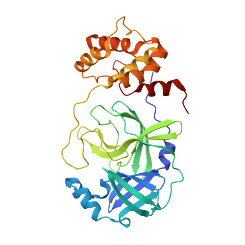
Structural Basis of the Main Proteases of Coronavirus Bound to Drug Candidate PF-07321332.
Li, J., Lin, C., Zhou, X., Zhong, F., Zeng, P., Yang, Y., Zhang, Y., Yu, B., Fan, X., McCormick, P.J., Fu, R., Fu, Y., Jiang, H., Zhang, J.(2022) J Virol 96: e0201321-e0201321
- PubMed: 35389231
- DOI: https://doi.org/10.1128/jvi.02013-21
- Primary Citation of Related Structures:
7VLO, 7VLP, 7VLQ, 7VTC - PubMed Abstract:
The high mutation rate of COVID-19 and the prevalence of multiple variants strongly support the need for pharmacological options to complement vaccine strategies. One region that appears highly conserved among different genera of coronaviruses is the substrate-binding site of the main protease (M pro or 3CL pro ), making it an attractive target for the development of broad-spectrum drugs for multiple coronaviruses. PF-07321332, developed by Pfizer, is the first orally administered inhibitor targeting the main protease of SARS-CoV-2, which also has shown potency against other coronaviruses. Here, we report three crystal structures of the main protease of SARS-CoV-2, SARS-CoV, and Middle East respiratory syndrome (MERS)-CoV bound to the inhibitor PF-07321332. The structures reveal a ligand-binding site that is conserved among SARS-CoV-2, SARS-CoV, and MERS-CoV, providing insights into the mechanism of inhibition of viral replication. The long and narrow cavity in the cleft between domains I and II of the main protease harbors multiple inhibitor-binding sites, where PF-07321332 occupies subsites S1, S2, and S4 and appears more restricted than other inhibitors. A detailed analysis of these structures illuminated key structural determinants essential for inhibition and elucidated the binding mode of action of the main proteases from different coronaviruses. Given the importance of the main protease for the treatment of SARS-CoV-2 infection, insights derived from this study should accelerate the design of safer and more effective antivirals. IMPORTANCE The current pandemic of multiple variants has created an urgent need for effective inhibitors of SARS-CoV-2 to complement vaccine strategies. PF-07321332, developed by Pfizer, is the first orally administered coronavirus-specific main protease inhibitor approved by the FDA. We solved the crystal structures of the main protease of SARS-CoV-2, SARS-CoV, and MERS-CoV that bound to the PF-07321332, suggesting PF-07321332 is a broad-spectrum inhibitor for coronaviruses. Structures of the main protease inhibitor complexes present an opportunity to discover safer and more effective inhibitors for COVID-19.
Organizational Affiliation:
College of Pharmaceutical Sciences, Gannan Medical Universitygrid.440714.2, Ganzhou, China.