7VEK

Crystal structure of Phytolacca americana UGT3 with capsaicin and UDP-2fluoroglucose


Experimental Data Snapshot

  • Method: X-RAY DIFFRACTION
  • Resolution: 2.60 Å
  • R-Value Free: 0.258 
  • R-Value Work: 0.204 
  • R-Value Observed: 0.207 

wwPDB Validation   3D Report Full Report


Ligand Structure Quality Assessment 


This is version 1.2 of the entry. See complete history


Literature

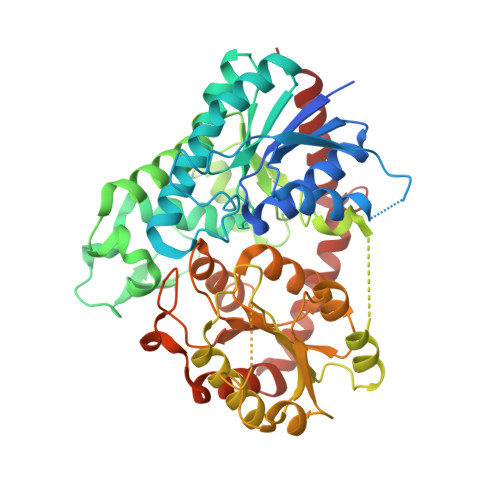
Structural basis for substrate recognition in the Phytolacca americana glycosyltransferase PaGT3.

Maharjan, R.Fukuda, Y.Nakayama, T.Nakayama, T.Hamada, H.Ozaki, S.I.Inoue, T.

(2022) Acta Crystallogr D Struct Biol 78: 379-389

  • DOI: https://doi.org/10.1107/S2059798322000869
  • Primary Citation of Related Structures:  
    7VEJ, 7VEK, 7VEL

  • PubMed Abstract: 

    Capsaicinoids are phenolic compounds that have health benefits. However, the pungency and poor water solubility of these compounds limit their exploitation. Glycosylation is a powerful method to improve water solubility and reduce pungency while preserving bioactivity. PaGT3, a uridine diphosphate glycosyltransferase (UGT) from Phytolacca americana, is known for its ability to glycosylate capsaicinoids and other phenolic compounds. While structural information on several UGTs is available, structures of UGTs that can glycosylate a range of phenolic compounds are rare. To fill this gap, crystal structures of PaGT3 with a sugar-donor analogue (UDP-2-fluoroglucose) and the acceptors capsaicin and kaempferol were determined. PaGT3 adopts a GT-B-fold structure that is highly conserved among UGTs. However, the acceptor-binding pocket in PaGT3 is hydrophobic and large, and is surrounded by longer loops. The larger acceptor-binding pocket in PaGT3 allows the enzyme to bind a range of compounds, while the flexibility of the longer loops possibly plays a role in accommodating the acceptors in the binding pocket according to their shape and size. This structural information provides insights into the acceptor-binding mechanism in UGTs that bind multiple substrates.


  • Organizational Affiliation

    Graduate School of Pharmaceutical Science, Osaka University, Suita, Osaka 565-0871, Japan.


Macromolecules
Find similar proteins by:  (by identity cutoff)  |  3D Structure
Entity ID: 1
MoleculeChains Sequence LengthOrganismDetailsImage
Glycosyltransferase
A, B
505Phytolacca americanaMutation(s): 0 
Gene Names: PaGT3
EC: 2.4.1
UniProt
Find proteins for B5MGN9 (Phytolacca americana)
Explore B5MGN9 
Go to UniProtKB:  B5MGN9
Entity Groups  
Sequence Clusters30% Identity50% Identity70% Identity90% Identity95% Identity100% Identity
UniProt GroupB5MGN9
Sequence Annotations
Expand
  • Reference Sequence
Small Molecules
Ligands 4 Unique
IDChains Name / Formula / InChI Key2D Diagram3D Interactions
U2F (Subject of Investigation/LOI)
Query on U2F

Download Ideal Coordinates CCD File 
E [auth A],
G [auth B]
URIDINE-5'-DIPHOSPHATE-2-DEOXY-2-FLUORO-ALPHA-D-GLUCOSE
C15 H23 F N2 O16 P2
NGTCPFGWXMBZEP-NQQHDEILSA-N
4DY (Subject of Investigation/LOI)
Query on 4DY

Download Ideal Coordinates CCD File 
F [auth B](6E)-N-(4-hydroxy-3-methoxybenzyl)-8-methylnon-6-enamide
C18 H27 N O3
YKPUWZUDDOIDPM-SOFGYWHQSA-N
O4B
Query on O4B

Download Ideal Coordinates CCD File 
C [auth A]1,4,7,10,13,16-HEXAOXACYCLOOCTADECANE
C12 H24 O6
XEZNGIUYQVAUSS-UHFFFAOYSA-N
K
Query on K

Download Ideal Coordinates CCD File 
D [auth A]POTASSIUM ION
K
NPYPAHLBTDXSSS-UHFFFAOYSA-N
Experimental Data & Validation

Experimental Data

  • Method: X-RAY DIFFRACTION
  • Resolution: 2.60 Å
  • R-Value Free: 0.258 
  • R-Value Work: 0.204 
  • R-Value Observed: 0.207 
  • Space Group: P 21 21 21
Unit Cell:
Length ( Å )Angle ( ˚ )
a = 94.276α = 90
b = 103.022β = 90
c = 109.648γ = 90
Software Package:
Software NamePurpose
PHENIXrefinement
XDSdata reduction
Aimlessdata scaling
MOLREPphasing

Structure Validation

View Full Validation Report


Ligand Structure Quality Assessment 


Entry History & Funding Information

Deposition Data


Funding OrganizationLocationGrant Number
Japan Society for the Promotion of Science (JSPS)Japan--

Revision History  (Full details and data files)

  • Version 1.0: 2022-03-02
    Type: Initial release
  • Version 1.1: 2022-03-16
    Changes: Database references
  • Version 1.2: 2023-11-29
    Changes: Data collection, Refinement description