7U6A

Crystal Structure of Danio rerio Histone Deacetylase 10 in Complex with 3-thienylmethyl Benzhydroxamic Acid Inhibitor


Experimental Data Snapshot

  • Method: X-RAY DIFFRACTION
  • Resolution: 2.25 Å
  • R-Value Free: 0.227 
  • R-Value Work: 0.195 
  • R-Value Observed: 0.197 

wwPDB Validation   3D Report Full Report


Ligand Structure Quality Assessment 


This is version 1.2 of the entry. See complete history


Literature

Identification of histone deacetylase 10 (HDAC10) inhibitors that modulate autophagy in transformed cells.

Zeyen, P.Zeyn, Y.Herp, D.Mahmoudi, F.Yesiloglu, T.Z.Erdmann, F.Schmidt, M.Robaa, D.Romier, C.Ridinger, J.Herbst-Gervasoni, C.J.Christianson, D.W.Oehme, I.Jung, M.Kramer, O.H.Sippl, W.

(2022) Eur J Med Chem 234: 114272-114272

  • DOI: https://doi.org/10.1016/j.ejmech.2022.114272
  • Primary Citation of Related Structures:  
    7U3M, 7U69, 7U6A, 7U6B

  • PubMed Abstract: 

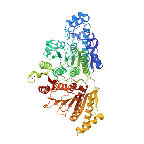
    Histone deacetylases (HDACs) are a family of 18 epigenetic modifiers that fall into 4 classes. Histone deacetylase inhibitors (HDACi) are valid tools to assess HDAC functions. HDAC6 and HDAC10 belong to the class IIb subgroup of the HDAC family. The targets and biological functions of HDAC10 are ill-defined. This lack of knowledge is due to a lack of specific and potent HDAC10 inhibitors with cellular activity. Here, we have synthesized and characterized piperidine-4-acrylhydroxamates as potent and highly selective inhibitors of HDAC10. This was achieved by targeting the acidic gatekeeper residue Glu274 of HDAC10 with a basic piperidine moiety that mimics the interaction of the polyamine substrate of HDAC10. We have confirmed the binding modes of selected inhibitors using X-ray crystallography. Promising candidates were selected based on their specificity by in vitro profiling using recombinant HDACs. The most promising HDAC10 inhibitors 10c and 13b were tested for specificity in acute myeloid leukemia (AML) cells with the FLT3-ITD oncogene. By immunoblot experiments we assessed the hyperacetylation of histones and tubulin-α, which are class I and HDAC6 substrates, respectively. As validated test for HDAC10 inhibition we used flow cytometry assessing autolysosome formation in neuroblastoma and AML cells. We demonstrate that 10c and 13b inhibit HDAC10 with high specificity over HDAC6 and with no significant impact on class I HDACs. The accumulation of autolysosomes is not a consequence of apoptosis and 10c and 13b are not toxic for normal human kidney cells. These data show that 10c and 13b are nanomolar inhibitors of HDAC10 with high specificity. Thus, our new HDAC10 inhibitors are tools to identify the downstream targets and functions of HDAC10 in cells.


  • Organizational Affiliation

    Institute of Pharmacy, Martin-Luther University of Halle-Wittenberg, Wolfgang-Langenbeck-Str. 2-4, 06120, Halle/Saale, Germany.


Macromolecules
Find similar proteins by:  (by identity cutoff)  |  3D Structure
Entity ID: 1
MoleculeChains Sequence LengthOrganismDetailsImage
Polyamine deacetylase HDAC10676Danio rerioMutation(s): 0 
Gene Names: hdac10
EC: 3.5.1.48 (PDB Primary Data), 3.5.1.62 (PDB Primary Data)
UniProt
Find proteins for F1QCV2 (Danio rerio)
Explore F1QCV2 
Go to UniProtKB:  F1QCV2
Entity Groups  
Sequence Clusters30% Identity50% Identity70% Identity90% Identity95% Identity100% Identity
UniProt GroupF1QCV2
Sequence Annotations
Expand
  • Reference Sequence
Small Molecules
Ligands 5 Unique
IDChains Name / Formula / InChI Key2D Diagram3D Interactions
LSL (Subject of Investigation/LOI)
Query on LSL

Download Ideal Coordinates CCD File 
B [auth A]N-hydroxy-4-({[(thiophen-3-yl)methyl]amino}methyl)benzamide
C13 H14 N2 O2 S
UUYNWXQYZUGELG-UHFFFAOYSA-N
PO4
Query on PO4

Download Ideal Coordinates CCD File 
G [auth A],
H [auth A],
I [auth A]
PHOSPHATE ION
O4 P
NBIIXXVUZAFLBC-UHFFFAOYSA-K
ZN
Query on ZN

Download Ideal Coordinates CCD File 
F [auth A]ZINC ION
Zn
PTFCDOFLOPIGGS-UHFFFAOYSA-N
EDO
Query on EDO

Download Ideal Coordinates CCD File 
C [auth A]1,2-ETHANEDIOL
C2 H6 O2
LYCAIKOWRPUZTN-UHFFFAOYSA-N
K
Query on K

Download Ideal Coordinates CCD File 
D [auth A],
E [auth A]
POTASSIUM ION
K
NPYPAHLBTDXSSS-UHFFFAOYSA-N
Experimental Data & Validation

Experimental Data

  • Method: X-RAY DIFFRACTION
  • Resolution: 2.25 Å
  • R-Value Free: 0.227 
  • R-Value Work: 0.195 
  • R-Value Observed: 0.197 
  • Space Group: P 31 2 1
Unit Cell:
Length ( Å )Angle ( ˚ )
a = 80.748α = 90
b = 80.748β = 90
c = 248.919γ = 120
Software Package:
Software NamePurpose
PHENIXrefinement
iMOSFLMdata reduction
Aimlessdata scaling
PHASERphasing

Structure Validation

View Full Validation Report


Ligand Structure Quality Assessment 


Entry History & Funding Information

Deposition Data


Funding OrganizationLocationGrant Number
National Institutes of Health/National Human Genome Research Institute (NIH/NHGRI)United StatesGM49758

Revision History  (Full details and data files)

  • Version 1.0: 2022-04-06
    Type: Initial release
  • Version 1.1: 2022-06-15
    Changes: Database references
  • Version 1.2: 2023-10-18
    Changes: Data collection, Refinement description