7U59

Crystal Structure of Danio rerio Histone Deacetylase 10 in Complex with Piperidine-4-hydroxamic acid Inhibitor


Experimental Data Snapshot

  • Method: X-RAY DIFFRACTION
  • Resolution: 2.18 Å
  • R-Value Free: 0.210 
  • R-Value Work: 0.179 
  • R-Value Observed: 0.181 

wwPDB Validation   3D Report Full Report


Ligand Structure Quality Assessment 


This is version 1.1 of the entry. See complete history


Literature

First Fluorescent Acetylspermidine Deacetylation Assay for HDAC10 Identifies Selective Inhibitors with Cellular Target Engagement.

Herp, D.Ridinger, J.Robaa, D.Shinsky, S.A.Schmidtkunz, K.Yesiloglu, T.Z.Bayer, T.Steimbach, R.R.Herbst-Gervasoni, C.J.Merz, A.Romier, C.Sehr, P.Gunkel, N.Miller, A.K.Christianson, D.W.Oehme, I.Sippl, W.Jung, M.

(2022) Chembiochem 23: e202200180-e202200180

  • DOI: https://doi.org/10.1002/cbic.202200180
  • Primary Citation of Related Structures:  
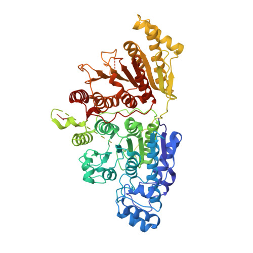
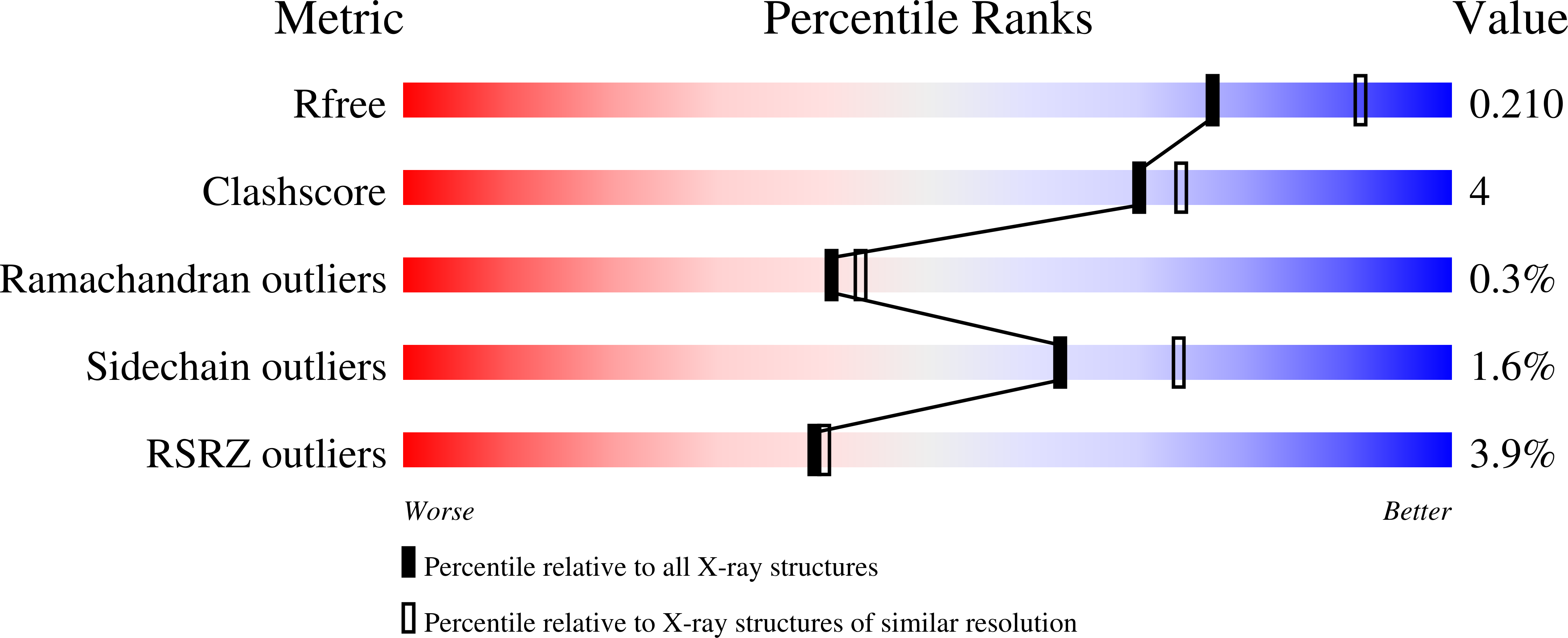
    7U59

  • PubMed Abstract: 

    Histone deacetylases (HDACs) are important epigenetic regulators involved in many diseases, especially cancer. Five HDAC inhibitors have been approved for anticancer therapy and many are in clinical trials. Among the 11 zinc-dependent HDACs, HDAC10 has received relatively little attention by drug discovery campaigns, despite its involvement, e. g., in the pathogenesis of neuroblastoma. This is due in part to a lack of robust enzymatic conversion assays. In contrast to the protein lysine deacetylase and deacylase activity of most other HDAC subtypes, it has recently been shown that HDAC10 has strong preferences for deacetylation of oligoamine substrates like acetyl-putrescine or -spermidine. Hence, it is also termed a polyamine deacetylase (PDAC). Here, we present the first fluorescent enzymatic conversion assay for HDAC10 using an aminocoumarin-labelled acetyl-spermidine derivative to measure its PDAC activity, which is suitable for high-throughput screening. Using this assay, we identified potent inhibitors of HDAC10-mediated spermidine deacetylation in vitro. Based on the oligoamine preference of HDAC10, we also designed inhibitors with a basic moiety in appropriate distance to the zinc binding hydroxamate that showed potent inhibition of HDAC10 with high selectivity, and we solved a HDAC10-inhibitor structure using X-ray crystallography. We could demonstrate selective cellular target engagement for HDAC10 but a lysosomal phenotype in neuroblastoma cells that was previously associated with HDAC10 inhibition was not observed. Thus, we have developed new chemical probes for HDAC10 that allow further clarification of the biological role of this enzyme.


  • Organizational Affiliation

    Institute of Pharmaceutical Sciences, University of Freiburg, Albertstraße 25, 79104, Freiburg, Germany.


Macromolecules
Find similar proteins by:  (by identity cutoff)  |  3D Structure
Entity ID: 1
MoleculeChains Sequence LengthOrganismDetailsImage
Polyamine deacetylase HDAC10676Danio rerioMutation(s): 0 
Gene Names: hdac10
EC: 3.5.1.48 (PDB Primary Data), 3.5.1.62 (PDB Primary Data)
UniProt
Find proteins for F1QCV2 (Danio rerio)
Explore F1QCV2 
Go to UniProtKB:  F1QCV2
Entity Groups  
Sequence Clusters30% Identity50% Identity70% Identity90% Identity95% Identity100% Identity
UniProt GroupF1QCV2
Sequence Annotations
Expand
  • Reference Sequence
Small Molecules
Ligands 6 Unique
IDChains Name / Formula / InChI Key2D Diagram3D Interactions
LDI (Subject of Investigation/LOI)
Query on LDI

Download Ideal Coordinates CCD File 
E [auth A]1-[2-(benzylamino)ethyl]-N-hydroxypiperidine-4-carboxamide
C15 H23 N3 O2
IKRHDIZZKCLEPC-UHFFFAOYSA-N
PO4
Query on PO4

Download Ideal Coordinates CCD File 
I [auth A],
J [auth A],
K [auth A]
PHOSPHATE ION
O4 P
NBIIXXVUZAFLBC-UHFFFAOYSA-K
ZN
Query on ZN

Download Ideal Coordinates CCD File 
F [auth A]ZINC ION
Zn
PTFCDOFLOPIGGS-UHFFFAOYSA-N
EDO
Query on EDO

Download Ideal Coordinates CCD File 
B [auth A],
C [auth A],
D [auth A]
1,2-ETHANEDIOL
C2 H6 O2
LYCAIKOWRPUZTN-UHFFFAOYSA-N
K
Query on K

Download Ideal Coordinates CCD File 
G [auth A],
H [auth A]
POTASSIUM ION
K
NPYPAHLBTDXSSS-UHFFFAOYSA-N
NA
Query on NA

Download Ideal Coordinates CCD File 
L [auth A]SODIUM ION
Na
FKNQFGJONOIPTF-UHFFFAOYSA-N
Experimental Data & Validation

Experimental Data

  • Method: X-RAY DIFFRACTION
  • Resolution: 2.18 Å
  • R-Value Free: 0.210 
  • R-Value Work: 0.179 
  • R-Value Observed: 0.181 
  • Space Group: P 31 2 1
Unit Cell:
Length ( Å )Angle ( ˚ )
a = 80.849α = 90
b = 80.849β = 90
c = 247.467γ = 120
Software Package:
Software NamePurpose
PHENIXrefinement
iMOSFLMdata reduction
Aimlessdata scaling
PHASERphasing

Structure Validation

View Full Validation Report


Ligand Structure Quality Assessment 


Entry History & Funding Information

Deposition Data


Funding OrganizationLocationGrant Number
National Institutes of Health/National Human Genome Research Institute (NIH/NHGRI)United StatesGM49758

Revision History  (Full details and data files)

  • Version 1.0: 2023-01-11
    Type: Initial release
  • Version 1.1: 2023-10-25
    Changes: Data collection, Refinement description