Functional aspects of evolution in a cluster of salivary protein genes from mosquitoes.
Alvarenga, P.H., Dias, D.R., Xu, X., Francischetti, I.M.B., Gittis, A.G., Arp, G., Garboczi, D.N., Ribeiro, J.M.C., Andersen, J.F.(2022) Insect Biochem Mol Biol 146: 103785-103785
- PubMed: 35568118
- DOI: https://doi.org/10.1016/j.ibmb.2022.103785
- Primary Citation of Related Structures:
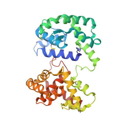
7TVC, 7TVY, 7TX8, 7U1N - PubMed Abstract:
The D7 proteins are highly expressed in the saliva of hematophagous Nematocera and bind biogenic amines and eicosanoid compounds produced by the host during blood feeding. These proteins are encoded by gene clusters expressing forms having one or two odorant-binding protein-like domains. Here we examine functional diversity within the D7 group in the genus Anopheles and make structural comparisons with D7 proteins from culicine mosquitoes in order to understand aspects of D7 functional evolution. Two domain long form (D7L) and one domain short form (D7S) proteins from anopheline and culicine mosquitoes were characterized to determine their ligand selectivity and binding pocket structures. We previously showed that a D7L protein from Anopheles stephensi, of the subgenus Cellia, could bind eicosanoids at a site in its N-terminal domain but could not bind biogenic amines in its C-terminal domain as does a D7L1 ortholog from the culicine species Aedes aegypti, raising the question of whether anopheline D7L proteins had lost their ability to bind biogenic amines. Here we find that D7L from anopheline species belonging to two other subgenera, Nyssorhynchus and Anopheles, can bind biogenic amines and have a structure much like the Ae. aegypti ortholog. The unusual D7L, D7L3, can also bind serotonin in the Cellia species An. gambiae. We also show through structural comparisons with culicine forms that the biogenic amine binding function of single domain D7S proteins in the genus Anopheles may have evolved through gene conversion of structurally similar proteins, which did not have biogenic amine binding capability. Collectively, the data indicate that D7L proteins had a biogenic amine and eicosanoid binding function in the common ancestor of anopheline and culicine mosquitoes, and that the D7S proteins may have acquired a biogenic amine binding function in anophelines through a gene conversion process.
Organizational Affiliation:
Laboratory of Malaria and Vector Research, National Institutes of Health, National Institute of Allergy and Infectious Diseases, Rockville, MD, 20852, USA; Laboratório de Bioquímica de Resposta ao Estresse, Instituto de Bioquímica Médica, Universidade Federal do Rio de Janeiro, Rio de Janeiro, 21941-902, Brazil. Electronic address: patricia.alvarenga@nih.gov.