


Structure of myxoma virus M062 protein variant MAV
O'Byrne, P., Khan, A.R.To be published.
Experimental Data Snapshot
wwPDB Validation 3D Report Full Report

Entity ID: 1 | |||||
|---|---|---|---|---|---|
| Molecule | Chains | Sequence Length | Organism | Details | Image |
| Probable host range protein 2-1 | 161 | Myxoma virus (strain Lausanne) | Mutation(s): 3 Gene Names: m062R | 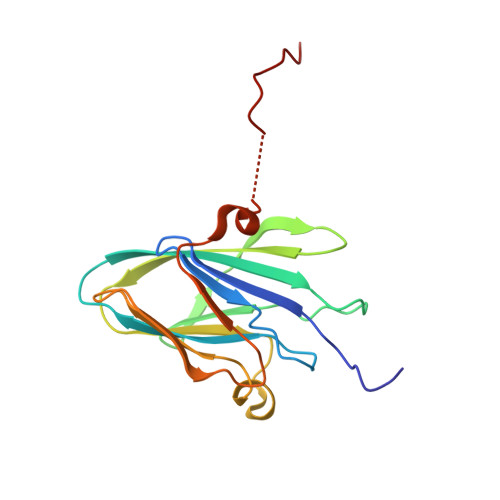 | |
UniProt | |||||
Find proteins for P68550 (Myxoma virus (strain Lausanne)) Explore P68550 Go to UniProtKB: P68550 | |||||
Entity Groups | |||||
| Sequence Clusters | 30% Identity50% Identity70% Identity90% Identity95% Identity100% Identity | ||||
| UniProt Group | P68550 | ||||
Sequence AnnotationsExpand | |||||
| |||||
| Ligands 1 Unique | |||||
|---|---|---|---|---|---|
| ID | Chains | Name / Formula / InChI Key | 2D Diagram | 3D Interactions | |
| NI Query on NI | B [auth A] | NICKEL (II) ION Ni VEQPNABPJHWNSG-UHFFFAOYSA-N |  | ||
| Length ( Å ) | Angle ( ˚ ) |
|---|---|
| a = 94.827 | α = 90 |
| b = 94.827 | β = 90 |
| c = 44.136 | γ = 120 |
| Software Name | Purpose |
|---|---|
| PHENIX | refinement |
| PDB_EXTRACT | data extraction |
| XDS | data reduction |
| Aimless | data scaling |
| PHASER | phasing |
| Funding Organization | Location | Grant Number |
|---|---|---|
| Science Foundation Ireland | Ireland | SFI 12/1A/1239 |
RCSB PDB (citation) is hosted by
RCSB PDB is a member of the

