7U0Q

SARS-Cov2 S protein structure in complex with neutralizing monoclonal antibody 002-02


Experimental Data Snapshot

  • Method: ELECTRON MICROSCOPY
  • Resolution: 3.86 Å
  • Aggregation State: PARTICLE 
  • Reconstruction Method: SINGLE PARTICLE 

wwPDB Validation   3D Report Full Report


This is version 1.1 of the entry. See complete history


Literature

Molecular basis of SARS-CoV-2 Omicron variant evasion from shared neutralizing antibody response.

Patel, A.Kumar, S.Lai, L.Chakravarthy, C.Valanparambil, R.Reddy, E.S.Gottimukkala, K.Bajpai, P.Raju, D.R.Edara, V.V.Davis-Gardner, M.E.Linderman, S.Dixit, K.Sharma, P.Mantus, G.Cheedarla, N.Verkerke, H.P.Frank, F.Neish, A.S.Roback, J.D.Davis, C.W.Wrammert, J.Ahmed, R.Suthar, M.S.Sharma, A.Murali-Krishna, K.Chandele, A.Ortlund, E.A.

(2023) Structure 31: 801-811.e5

  • DOI: https://doi.org/10.1016/j.str.2023.04.010
  • Primary Citation of Related Structures:  
    7U0Q, 7U0X, 7UOW

  • PubMed Abstract: 

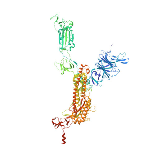
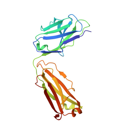
    Understanding the molecular features of neutralizing epitopes is important for developing vaccines/therapeutics against emerging SARS-CoV-2 variants. We describe three monoclonal antibodies (mAbs) generated from COVID-19 recovered individuals during the first wave of the pandemic in India. These mAbs had publicly shared near germline gene usage and potently neutralized Alpha and Delta, poorly neutralized Beta, and failed to neutralize Omicron BA.1 SARS-CoV-2 variants. Structural analysis of these mAbs in complex with trimeric spike protein showed that all three mAbs bivalently bind spike with two mAbs targeting class 1 and one targeting a class 4 receptor binding domain epitope. The immunogenetic makeup, structure, and function of these mAbs revealed specific molecular interactions associated with the potent multi-variant binding/neutralization efficacy. This knowledge shows how mutational combinations can affect the binding or neutralization of an antibody, which in turn relates to the efficacy of immune responses to emerging SARS-CoV-2 escape variants.


  • Organizational Affiliation

    Department of Biochemistry, Emory University School of Medicine, Atlanta, GA 30322, USA.


Macromolecules
Find similar proteins by:  (by identity cutoff)  |  3D Structure
Entity ID: 1
MoleculeChains Sequence LengthOrganismDetailsImage
Spike glycoprotein
A, B, C
1,208Severe acute respiratory syndrome coronavirus 2Mutation(s): 0 
Gene Names: S2
UniProt
Find proteins for P0DTC2 (Severe acute respiratory syndrome coronavirus 2)
Explore P0DTC2 
Go to UniProtKB:  P0DTC2
Entity Groups  
Sequence Clusters30% Identity50% Identity70% Identity90% Identity95% Identity100% Identity
UniProt GroupP0DTC2
Sequence Annotations
Expand
  • Reference Sequence
Find similar proteins by:  (by identity cutoff)  |  3D Structure
Entity ID: 2
MoleculeChains Sequence LengthOrganismDetailsImage
mAb 002-02 light chainD [auth F],
F [auth I]
214Homo sapiensMutation(s): 0 
Entity Groups  
Sequence Clusters30% Identity50% Identity70% Identity90% Identity95% Identity100% Identity
Sequence Annotations
Expand
  • Reference Sequence
Find similar proteins by:  (by identity cutoff)  |  3D Structure
Entity ID: 3
MoleculeChains Sequence LengthOrganismDetailsImage
mAb 002-02 Heavy chainE [auth G],
G [auth J]
447Homo sapiensMutation(s): 0 
Entity Groups  
Sequence Clusters30% Identity50% Identity70% Identity90% Identity95% Identity100% Identity
Sequence Annotations
Expand
  • Reference Sequence
Oligosaccharides

Help

Entity ID: 4
MoleculeChains Length2D Diagram Glycosylation3D Interactions
2-acetamido-2-deoxy-beta-D-glucopyranose-(1-4)-2-acetamido-2-deoxy-beta-D-glucopyranose
H [auth D],
I [auth E],
J [auth H],
K,
L,
H [auth D],
I [auth E],
J [auth H],
K,
L,
M,
N,
O,
P,
Q,
R,
S,
T,
U
2N-Glycosylation
Glycosylation Resources
GlyTouCan:  G42666HT
GlyCosmos:  G42666HT
GlyGen:  G42666HT
Entity ID: 5
MoleculeChains Length2D Diagram Glycosylation3D Interactions
beta-D-mannopyranose-(1-4)-2-acetamido-2-deoxy-beta-D-glucopyranose-(1-4)-2-acetamido-2-deoxy-beta-D-glucopyranose
V
3N-Glycosylation
Glycosylation Resources
GlyTouCan:  G15407YE
GlyCosmos:  G15407YE
GlyGen:  G15407YE
Small Molecules
Ligands 1 Unique
IDChains Name / Formula / InChI Key2D Diagram3D Interactions
NAG (Subject of Investigation/LOI)
Query on NAG

Download Ideal Coordinates CCD File 
AA [auth A]
BA [auth A]
CA [auth A]
DA [auth A]
EA [auth A]
AA [auth A],
BA [auth A],
CA [auth A],
DA [auth A],
EA [auth A],
FA [auth B],
GA [auth B],
HA [auth B],
IA [auth B],
JA [auth B],
KA [auth B],
LA [auth B],
MA [auth C],
NA [auth C],
OA [auth C],
PA [auth C],
QA [auth C],
RA [auth C],
SA [auth C],
W [auth A],
X [auth A],
Y [auth A],
Z [auth A]
2-acetamido-2-deoxy-beta-D-glucopyranose
C8 H15 N O6
OVRNDRQMDRJTHS-FMDGEEDCSA-N
Experimental Data & Validation

Experimental Data

  • Method: ELECTRON MICROSCOPY
  • Resolution: 3.86 Å
  • Aggregation State: PARTICLE 
  • Reconstruction Method: SINGLE PARTICLE 
EM Software:
TaskSoftware PackageVersion
RECONSTRUCTIONcryoSPARC3.2.0
MODEL REFINEMENTPHENIX

Structure Validation

View Full Validation Report


Entry History & Funding Information

Deposition Data


Funding OrganizationLocationGrant Number
National Institutes of Health/National Institute of Biomedical Imaging and Bioengineering (NIH/NIBIB)United States3U54EB027690-04S

Revision History  (Full details and data files)

  • Version 1.0: 2023-03-01
    Type: Initial release
  • Version 1.1: 2023-09-13
    Changes: Data collection, Database references