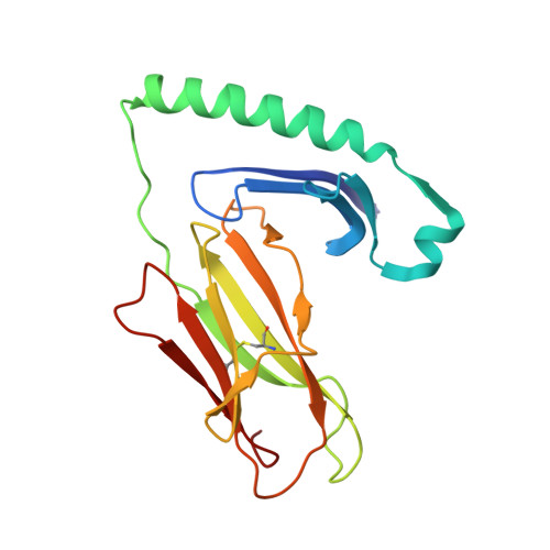
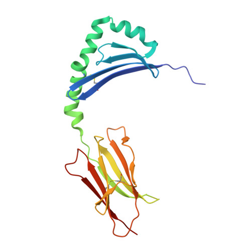
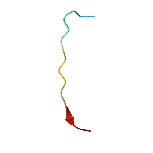
7T6I

Crystal structure of HLA-DP1 in complex with pp65 peptide in reverse orientation


Experimental Data Snapshot

  • Method: X-RAY DIFFRACTION
  • Resolution: 2.30 Å
  • R-Value Free: 0.224 
  • R-Value Work: 0.181 
  • R-Value Observed: 0.183 

wwPDB Validation   3D Report Full Report


Ligand Structure Quality Assessment 


This is version 1.1 of the entry. See complete history


Literature

Human T cells recognize HLA-DP-bound peptides in two orientations.

Klobuch, S.Lim, J.J.van Balen, P.Kester, M.G.D.de Klerk, W.de Ru, A.H.Pothast, C.R.Jedema, I.Drijfhout, J.W.Rossjohn, J.Reid, H.H.van Veelen, P.A.Falkenburg, J.H.F.Heemskerk, M.H.M.

(2022) Proc Natl Acad Sci U S A 119: e2214331119-e2214331119

  • DOI: https://doi.org/10.1073/pnas.2214331119
  • Primary Citation of Related Structures:  
    7T6I

  • PubMed Abstract: 

    Human leukocyte antigen (HLA) molecules present small peptide antigens to T cells, thereby allowing them to recognize pathogen-infected and cancer cells. A central dogma over the last 50+ y is that peptide binding to HLA molecules is mediated by the docking of side chains of particular amino acids in the peptide into pockets in the HLA molecules in a conserved N- to C-terminal orientation. Whether peptides can be presented in a reversed C- to N-terminal orientation remains unclear. Here, we performed large-scale identification of peptides bound to HLA-DP molecules and observed that in addition to peptide binding in an N- to C-terminal orientation, in 9 out of 14 HLA-DP allotypes, reverse motifs are found, compatible with C- to N-terminal peptide binding. Moreover, we isolated high-avidity human cytomegalovirus (CMV)-specific HLA-DP-restricted CD4 + T cells from the memory repertoire of healthy donors and demonstrate that such T cells recognized CMV-derived peptides bound to HLA-DPB1*01:01 or *05:01 in a reverse C- to N-terminal manner. Finally, we obtained a high-resolution HLA-DPB1*01:01-CMVpp65 (142-158) peptide crystal structure, which is the molecular basis for C- to N-terminal peptide binding to HLA-DP. Our results point to unique features of HLA-DP molecules that substantially broaden the HLA class II bound peptide repertoire to combat pathogens and eliminate cancer cells.


  • Organizational Affiliation

    Department of Hematology, Leiden University Medical Center, 2333 ZA Leiden, The Netherlands.


Macromolecules
Find similar proteins by:  (by identity cutoff)  |  3D Structure
Entity ID: 1
MoleculeChains Sequence LengthOrganismDetailsImage
HLA class II histocompatibility antigen DP alpha chain183Homo sapiensMutation(s): 0 
Gene Names: HLA-DPA1
UniProt
Find proteins for Q95HB9 (Homo sapiens)
Explore Q95HB9 
Go to UniProtKB:  Q95HB9
Entity Groups  
Sequence Clusters30% Identity50% Identity70% Identity90% Identity95% Identity100% Identity
UniProt GroupQ95HB9
Sequence Annotations
Expand
  • Reference Sequence
Find similar proteins by:  (by identity cutoff)  |  3D Structure
Entity ID: 2
MoleculeChains Sequence LengthOrganismDetailsImage
MHC class II antigen191Homo sapiensMutation(s): 0 
Gene Names: HLA-DPB1DPB1
UniProt
Find proteins for S6B6U4 (Homo sapiens)
Explore S6B6U4 
Go to UniProtKB:  S6B6U4
Entity Groups  
Sequence Clusters30% Identity50% Identity70% Identity90% Identity95% Identity100% Identity
UniProt GroupS6B6U4
Sequence Annotations
Expand
  • Reference Sequence

Find similar proteins by:  Sequence   |   3D Structure  

Entity ID: 3
MoleculeChains Sequence LengthOrganismDetailsImage
pp65 peptide17Human herpesvirus 5 strain AD169Mutation(s): 0 
UniProt
Find proteins for P06725 (Human cytomegalovirus (strain AD169))
Explore P06725 
Go to UniProtKB:  P06725
Entity Groups  
Sequence Clusters30% Identity50% Identity70% Identity90% Identity95% Identity100% Identity
UniProt GroupP06725
Sequence Annotations
Expand
  • Reference Sequence
Experimental Data & Validation

Experimental Data

  • Method: X-RAY DIFFRACTION
  • Resolution: 2.30 Å
  • R-Value Free: 0.224 
  • R-Value Work: 0.181 
  • R-Value Observed: 0.183 
  • Space Group: C 1 2 1
Unit Cell:
Length ( Å )Angle ( ˚ )
a = 112.36α = 90
b = 62.364β = 91.523
c = 72.796γ = 90
Software Package:
Software NamePurpose
PHENIXrefinement
XDSdata reduction
Aimlessdata scaling
PHASERphasing

Structure Validation

View Full Validation Report


Ligand Structure Quality Assessment 


Entry History & Funding Information

Deposition Data


Funding OrganizationLocationGrant Number
Australian Research Council (ARC)Australia--

Revision History  (Full details and data files)

  • Version 1.0: 2022-12-07
    Type: Initial release
  • Version 1.1: 2023-10-25
    Changes: Data collection, Refinement description