


Crystal Structure of Ebola zaire Envelope glycoprotein GP in complex with compound ARN0075093
Abendroth, J., Lorimer, D.D., Horanyi, P.S., Edwards, T.E.To be published.
Experimental Data Snapshot
Entity ID: 1 | |||||
|---|---|---|---|---|---|
| Molecule | Chains | Sequence Length | Organism | Details | Image |
| GP1 | 329 | Ebola virus - Mayinga, Zaire, 1976 | Mutation(s): 1 Gene Names: GP | 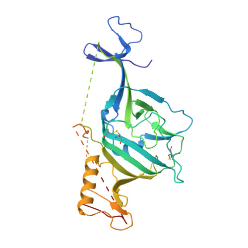 | |
UniProt | |||||
Find proteins for Q05320 (Zaire ebolavirus (strain Mayinga-76)) Explore Q05320 Go to UniProtKB: Q05320 | |||||
Entity Groups | |||||
| Sequence Clusters | 30% Identity50% Identity70% Identity90% Identity95% Identity100% Identity | ||||
| UniProt Group | Q05320 | ||||
Sequence AnnotationsExpand | |||||
| |||||
Entity ID: 2 | |||||
|---|---|---|---|---|---|
| Molecule | Chains | Sequence Length | Organism | Details | Image |
| GP2 | 168 | Ebola virus - Mayinga, Zaire, 1976 | Mutation(s): 1 Gene Names: GP |  | |
UniProt | |||||
Find proteins for Q05320 (Zaire ebolavirus (strain Mayinga-76)) Explore Q05320 Go to UniProtKB: Q05320 | |||||
Entity Groups | |||||
| Sequence Clusters | 30% Identity50% Identity70% Identity90% Identity95% Identity100% Identity | ||||
| UniProt Group | Q05320 | ||||
Sequence AnnotationsExpand | |||||
| |||||
Entity ID: 3 | |||||
|---|---|---|---|---|---|
| Molecule | Chains | Length | 2D Diagram | Glycosylation | 3D Interactions |
| alpha-D-mannopyranose-(1-6)-beta-D-mannopyranose-(1-4)-2-acetamido-2-deoxy-beta-D-glucopyranose-(1-4)-2-acetamido-2-deoxy-beta-D-glucopyranose | C | 4 |  | N-Glycosylation | |
Glycosylation Resources | |||||
GlyTouCan: G22573RC GlyCosmos: G22573RC GlyGen: G22573RC | |||||
| Ligands 4 Unique | |||||
|---|---|---|---|---|---|
| ID | Chains | Name / Formula / InChI Key | 2D Diagram | 3D Interactions | |
| ZTL (Subject of Investigation/LOI) Query on ZTL | K [auth B] | (1R,2s,3S,5s,7s)-N-[(1r,4r)-4-(aminomethyl)cyclohexyl]-5-phenyladamantane-2-carboxamide C24 H34 N2 O IRIFICPLNIURJD-IXFKACPXSA-N |  | ||
| NAG Query on NAG | D [auth A], E [auth A], F [auth A], G [auth A] | 2-acetamido-2-deoxy-beta-D-glucopyranose C8 H15 N O6 OVRNDRQMDRJTHS-FMDGEEDCSA-N |  | ||
| EDO Query on EDO | H [auth A] | 1,2-ETHANEDIOL C2 H6 O2 LYCAIKOWRPUZTN-UHFFFAOYSA-N |  | ||
| ACT Query on ACT | I [auth A], J [auth A] | ACETATE ION C2 H3 O2 QTBSBXVTEAMEQO-UHFFFAOYSA-M |  | ||
| Length ( Å ) | Angle ( ˚ ) |
|---|---|
| a = 114.07 | α = 90 |
| b = 114.07 | β = 90 |
| c = 308.09 | γ = 120 |
| Software Name | Purpose |
|---|---|
| XDS | data reduction |
| XSCALE | data scaling |
| PHENIX | refinement |
| PDB_EXTRACT | data extraction |
| PHASER | phasing |
| Funding Organization | Location | Grant Number |
|---|---|---|
| National Institutes of Health/National Institute Of Allergy and Infectious Diseases (NIH/NIAID) | United States | HHSN272201700059C |
RCSB PDB (citation) is hosted by
RCSB PDB is a member of the

