7SQX

Crystal Structure of Pseudomonas aeruginosa lytic polysaccharide monooxygenase CbpD


Experimental Data Snapshot

  • Method: X-RAY DIFFRACTION
  • Resolution: 3.00 Å
  • R-Value Free: 0.249 
  • R-Value Work: 0.210 
  • R-Value Observed: 0.213 

wwPDB Validation   3D Report Full Report


This is version 1.2 of the entry. See complete history


Literature

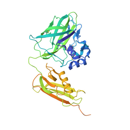
The crystal structure of CbpD clarifies substrate-specificity motifs in chitin-active lytic polysaccharide monooxygenases.

Dade, C.M.Douzi, B.Cambillau, C.Ball, G.Voulhoux, R.Forest, K.T.

(2022) Acta Crystallogr D Struct Biol 78: 1064-1078

  • DOI: https://doi.org/10.1107/S2059798322007033
  • Primary Citation of Related Structures:  
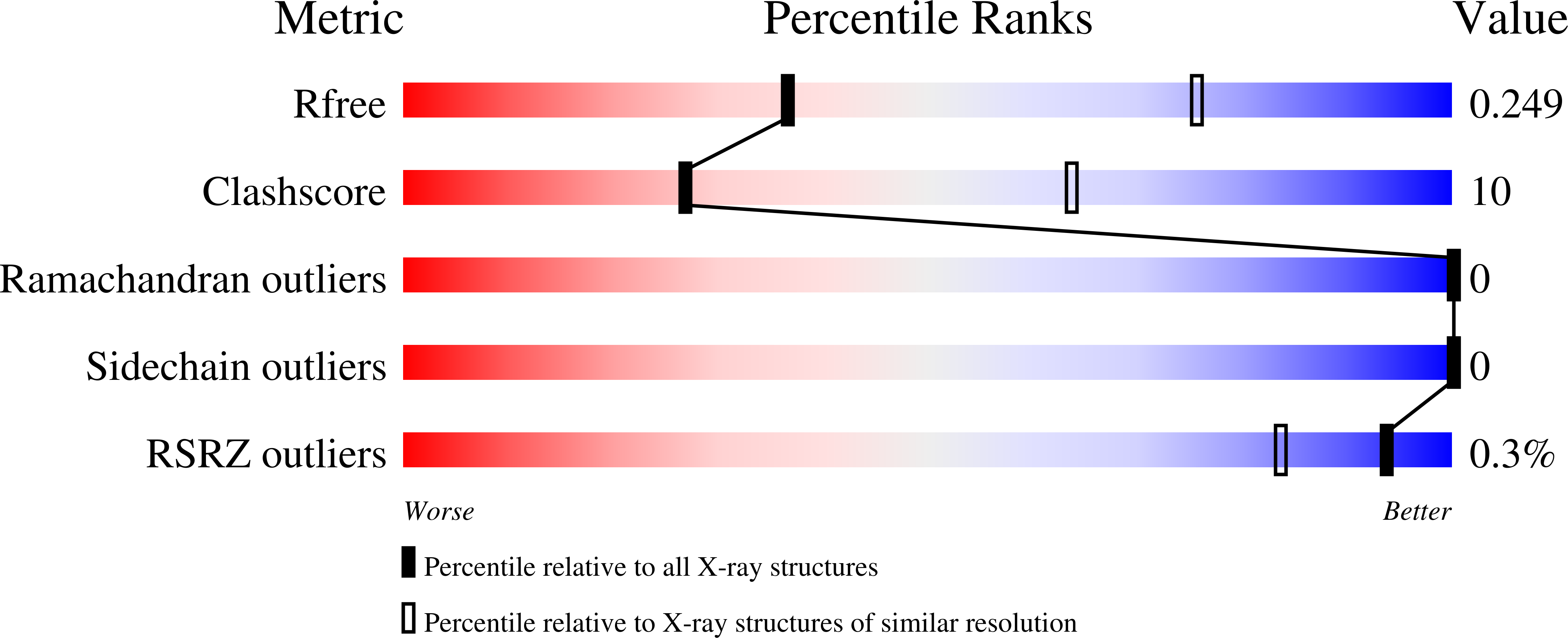
    7SQX

  • PubMed Abstract: 

    Pseudomonas aeruginosa secretes diverse proteins via its type 2 secretion system, including a 39 kDa chitin-binding protein, CbpD. CbpD has recently been shown to be a lytic polysaccharide monooxygenase active on chitin and to contribute substantially to virulence. To date, no structure of this virulence factor has been reported. Its first two domains are homologous to those found in the crystal structure of Vibrio cholerae GbpA, while the third domain is homologous to the NMR structure of the CBM73 domain of Cellvibrio japonicus CjLPMO10A. Here, the 3.0 Å resolution crystal structure of CbpD solved by molecular replacement is reported, which required ab initio models of each CbpD domain generated by the artificial intelligence deep-learning structure-prediction algorithm RoseTTAFold. The structure of CbpD confirms some previously reported substrate-specificity motifs among LPMOAA10s, while challenging the predictive power of others. Additionally, the structure of CbpD shows that post-translational modifications occur on the chitin-binding surface. Moreover, the structure raises interesting possibilities about how type 2 secretion-system substrates may interact with the secretion machinery and demonstrates the utility of new artificial intelligence protein structure-prediction algorithms in making challenging structural targets tractable.


  • Organizational Affiliation

    Department of Chemistry, University of Wisconsin-Madison, Madison, Wisconsin, USA.


Macromolecules
Find similar proteins by:  (by identity cutoff)  |  3D Structure
Entity ID: 1
MoleculeChains Sequence LengthOrganismDetailsImage
Chitin-binding protein CbpD374Pseudomonas aeruginosa PAO1Mutation(s): 0 
Gene Names: cbpDlasDPA0852
UniProt
Find proteins for Q9I589 (Pseudomonas aeruginosa (strain ATCC 15692 / DSM 22644 / CIP 104116 / JCM 14847 / LMG 12228 / 1C / PRS 101 / PAO1))
Explore Q9I589 
Go to UniProtKB:  Q9I589
Entity Groups  
Sequence Clusters30% Identity50% Identity70% Identity90% Identity95% Identity100% Identity
UniProt GroupQ9I589
Sequence Annotations
Expand
  • Reference Sequence
Experimental Data & Validation

Experimental Data

  • Method: X-RAY DIFFRACTION
  • Resolution: 3.00 Å
  • R-Value Free: 0.249 
  • R-Value Work: 0.210 
  • R-Value Observed: 0.213 
  • Space Group: P 31 1 2
Unit Cell:
Length ( Å )Angle ( ˚ )
a = 85.77α = 90
b = 85.77β = 90
c = 90.86γ = 120
Software Package:
Software NamePurpose
PHENIXrefinement
XSCALEdata scaling
PHASERphasing

Structure Validation

View Full Validation Report


Entry History & Funding Information

Deposition Data


Funding OrganizationLocationGrant Number
United States Department of Agriculture (USDA)FranceWIS03052
Agence Nationale de la Recherche (ANR)FranceANR-14-CE09-0027-01

Revision History  (Full details and data files)

  • Version 1.0: 2022-07-20
    Type: Initial release
  • Version 1.1: 2022-08-17
    Changes: Database references
  • Version 1.2: 2024-04-03
    Changes: Data collection, Refinement description