7SEZ

Crystal structure of Vaccinia Virus decapping enzyme D9 in complex with m7GDP


Experimental Data Snapshot

  • Method: X-RAY DIFFRACTION
  • Resolution: 1.70 Å
  • R-Value Free: 0.224 
  • R-Value Work: 0.193 
  • R-Value Observed: 0.195 

wwPDB Validation   3D Report Full Report


Ligand Structure Quality Assessment 


This is version 2.0 of the entry. See complete history


Literature

Structure of the poxvirus decapping enzyme D9 reveals its mechanism of cap recognition and catalysis.

Peters, J.K.Tibble, R.W.Warminski, M.Jemielity, J.Gross, J.D.

(2022) Structure 30: 721

  • DOI: https://doi.org/10.1016/j.str.2022.02.012
  • Primary Citation of Related Structures:  
    7SEZ, 7SF0

  • PubMed Abstract: 

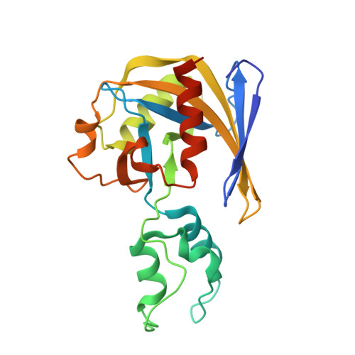
    Poxviruses encode decapping enzymes that remove the protective 5' cap from both host and viral mRNAs to commit transcripts for decay by the cellular exonuclease Xrn1. Decapping by these enzymes is critical for poxvirus pathogenicity by means of simultaneously suppressing host protein synthesis and limiting the accumulation of viral double-stranded RNA (dsRNA), a trigger for antiviral responses. Here we present a high-resolution structural view of the vaccinia virus decapping enzyme D9. This Nudix enzyme contains a domain organization different from other decapping enzymes in which a three-helix bundle is inserted into the catalytic Nudix domain. The 5' mRNA cap is positioned in a bipartite active site at the interface of the two domains. Specificity for the methylated guanosine cap is achieved by stacking between conserved aromatic residues in a manner similar to that observed in canonical cap-binding proteins VP39, eIF4E, and CBP20, and distinct from eukaryotic decapping enzyme Dcp2.


  • Organizational Affiliation

    Department of Pharmaceutical Chemistry, University of California, San Francisco, San Francisco, CA 94158, USA.


Macromolecules
Find similar proteins by:  (by identity cutoff)  |  3D Structure
Entity ID: 1
MoleculeChains Sequence LengthOrganismDetailsImage
DNA repair NTP-phosphohydrolase221Vaccinia virus Western ReserveMutation(s): 0 
Gene Names: 
UniProt
Find proteins for P04311 (Vaccinia virus (strain Western Reserve))
Explore P04311 
Go to UniProtKB:  P04311
Entity Groups  
Sequence Clusters30% Identity50% Identity70% Identity90% Identity95% Identity100% Identity
UniProt GroupP04311
Sequence Annotations
Expand
  • Reference Sequence
Small Molecules
Ligands 2 Unique
IDChains Name / Formula / InChI Key2D Diagram3D Interactions
M7G (Subject of Investigation/LOI)
Query on M7G

Download Ideal Coordinates CCD File 
B [auth A]7N-METHYL-8-HYDROGUANOSINE-5'-DIPHOSPHATE
C11 H18 N5 O11 P2
SBASPRRECYVBRF-KQYNXXCUSA-O
NA
Query on NA

Download Ideal Coordinates CCD File 
C [auth A]
D [auth A]
E [auth A]
F [auth A]
G [auth A]
C [auth A],
D [auth A],
E [auth A],
F [auth A],
G [auth A],
H [auth A],
I [auth A],
J [auth A],
K [auth A]
SODIUM ION
Na
FKNQFGJONOIPTF-UHFFFAOYSA-N
Experimental Data & Validation

Experimental Data

  • Method: X-RAY DIFFRACTION
  • Resolution: 1.70 Å
  • R-Value Free: 0.224 
  • R-Value Work: 0.193 
  • R-Value Observed: 0.195 
  • Space Group: C 2 2 21
Unit Cell:
Length ( Å )Angle ( ˚ )
a = 52.55α = 90
b = 140.1β = 90
c = 75.98γ = 90
Software Package:
Software NamePurpose
PHENIXrefinement
XDSdata reduction
XSCALEdata scaling
PHENIXphasing

Structure Validation

View Full Validation Report


Ligand Structure Quality Assessment 


Entry History & Funding Information

Deposition Data


Funding OrganizationLocationGrant Number
National Institutes of Health/National Institute of General Medical Sciences (NIH/NIGMS)United States2R01GM078360
National Institutes of Health/National Institute of General Medical Sciences (NIH/NIGMS)United States5F32GM133084

Revision History  (Full details and data files)

  • Version 1.0: 2022-03-30
    Type: Initial release
  • Version 1.1: 2022-05-18
    Changes: Database references
  • Version 2.0: 2024-02-28
    Changes: Atomic model, Author supporting evidence, Data collection, Derived calculations, Non-polymer description, Structure summary