7RNK

Room temperature X-ray structure of SARS-CoV-2 main protease (Mpro) in complex with HL-3-71


Experimental Data Snapshot

  • Method: X-RAY DIFFRACTION
  • Resolution: 2.10 Å
  • R-Value Free: 0.206 
  • R-Value Work: 0.176 
  • R-Value Observed: 0.177 

wwPDB Validation   3D Report Full Report


Ligand Structure Quality Assessment 


This is version 1.2 of the entry. See complete history


Literature

Structural, Electronic, and Electrostatic Determinants for Inhibitor Binding to Subsites S1 and S2 in SARS-CoV-2 Main Protease.

Kneller, D.W.Li, H.Galanie, S.Phillips, G.Labbe, A.Weiss, K.L.Zhang, Q.Arnould, M.A.Clyde, A.Ma, H.Ramanathan, A.Jonsson, C.B.Head, M.S.Coates, L.Louis, J.M.Bonnesen, P.V.Kovalevsky, A.

(2021) J Med Chem 64: 17366-17383

  • DOI: https://doi.org/10.1021/acs.jmedchem.1c01475
  • Primary Citation of Related Structures:  
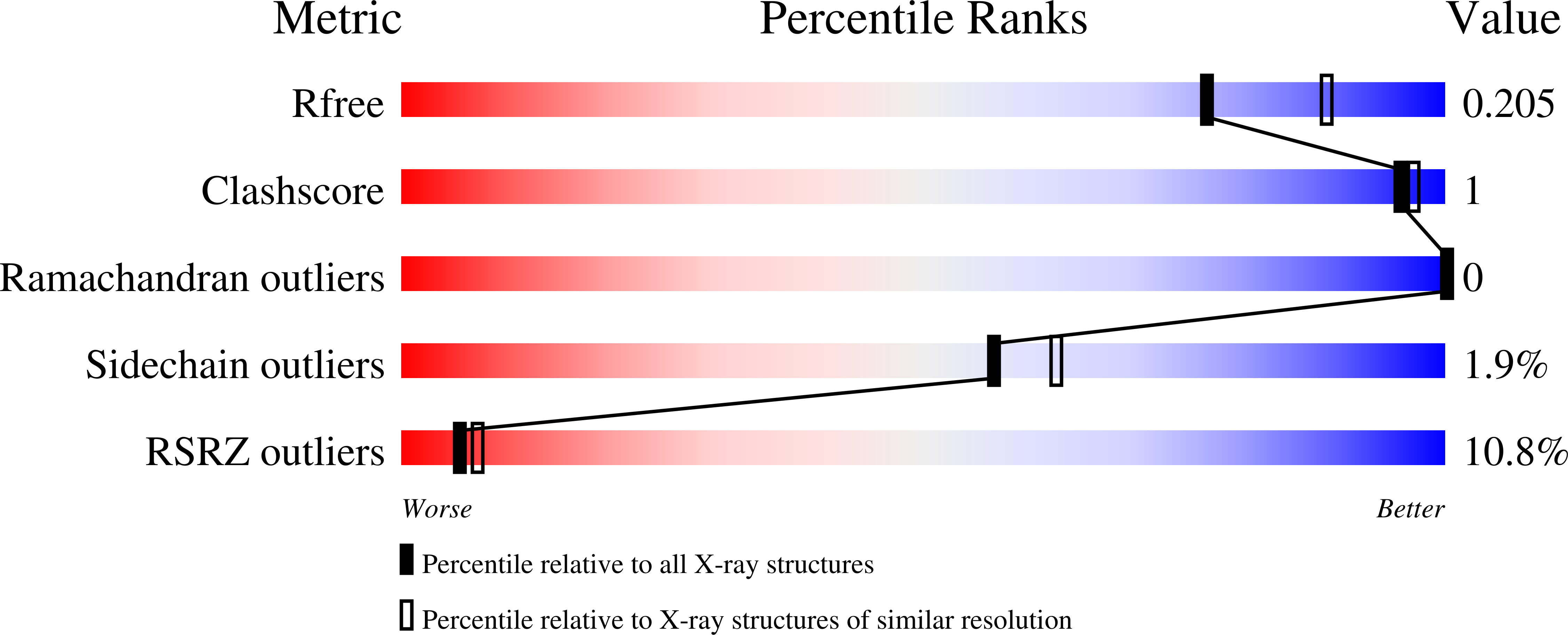
    7N8C, 7RLS, 7RM2, 7RMB, 7RME, 7RMT, 7RMZ, 7RN4, 7RNH, 7RNK

  • PubMed Abstract: 

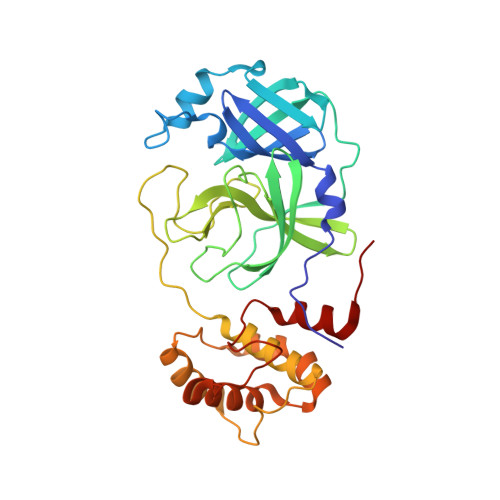
    Creating small-molecule antivirals specific for severe acute respiratory syndrome coronavirus 2 (SARS-CoV-2) proteins is crucial to battle coronavirus disease 2019 (COVID-19). SARS-CoV-2 main protease (M pro ) is an established drug target for the design of protease inhibitors. We performed a structure-activity relationship (SAR) study of noncovalent compounds that bind in the enzyme's substrate-binding subsites S1 and S2, revealing structural, electronic, and electrostatic determinants of these sites. The study was guided by the X-ray/neutron structure of M pro complexed with Mcule-5948770040 (compound 1 ), in which protonation states were directly visualized. Virtual reality-assisted structure analysis and small-molecule building were employed to generate analogues of 1 . In vitro enzyme inhibition assays and room-temperature X-ray structures demonstrated the effect of chemical modifications on M pro inhibition, showing that (1) maintaining correct geometry of an inhibitor's P1 group is essential to preserve the hydrogen bond with the protonated His163; (2) a positively charged linker is preferred; and (3) subsite S2 prefers nonbulky modestly electronegative groups.


  • Organizational Affiliation

    Neutron Scattering Division, Oak Ridge National Laboratory, Oak Ridge, Tennessee 37831, United States.


Macromolecules
Find similar proteins by:  (by identity cutoff)  |  3D Structure
Entity ID: 1
MoleculeChains Sequence LengthOrganismDetailsImage
3C-like proteinase306Severe acute respiratory syndrome coronavirus 2Mutation(s): 0 
Gene Names: rep1a-1b
EC: 3.4.22.69
UniProt
Find proteins for P0DTD1 (Severe acute respiratory syndrome coronavirus 2)
Explore P0DTD1 
Go to UniProtKB:  P0DTD1
Entity Groups  
Sequence Clusters30% Identity50% Identity70% Identity90% Identity95% Identity100% Identity
UniProt GroupP0DTD1
Sequence Annotations
Expand
  • Reference Sequence
Small Molecules
Ligands 1 Unique
IDChains Name / Formula / InChI Key2D Diagram3D Interactions
5ZT (Subject of Investigation/LOI)
Query on 5ZT

Download Ideal Coordinates CCD File 
B [auth A]6-{4-[3-chloro-4-(hydroxymethyl)phenyl]piperazine-1-carbonyl}pyrimidine-2,4(3H,5H)-dione
C16 H17 Cl N4 O4
LOQJVSKMCQMDKR-UHFFFAOYSA-N
Experimental Data & Validation

Experimental Data

  • Method: X-RAY DIFFRACTION
  • Resolution: 2.10 Å
  • R-Value Free: 0.206 
  • R-Value Work: 0.176 
  • R-Value Observed: 0.177 
  • Space Group: I 1 2 1
Unit Cell:
Length ( Å )Angle ( ˚ )
a = 52.284α = 90
b = 81.607β = 95.5
c = 91.432γ = 90
Software Package:
Software NamePurpose
PHENIXrefinement
CrysalisProdata reduction
Aimlessdata scaling
PHASERphasing

Structure Validation

View Full Validation Report


Ligand Structure Quality Assessment 


Entry History 

Deposition Data

Revision History  (Full details and data files)

  • Version 1.0: 2021-11-10
    Type: Initial release
  • Version 1.1: 2021-12-22
    Changes: Database references
  • Version 1.2: 2023-10-18
    Changes: Data collection, Refinement description