7QYS

Crystal structure of RimK from Pseudomonas syringae DC3000


Experimental Data Snapshot

  • Method: X-RAY DIFFRACTION
  • Resolution: 2.90 Å
  • R-Value Free: 0.248 
  • R-Value Work: 0.237 
  • R-Value Observed: 0.238 

wwPDB Validation   3D Report Full Report


Ligand Structure Quality Assessment 


This is version 1.2 of the entry. See complete history


Literature

Structural insights into the mechanism of adaptive ribosomal modification by Pseudomonas RimK.

Thompson, C.M.A.Little, R.H.Stevenson, C.E.M.Lawson, D.M.Malone, J.G.

(2023) Proteins 91: 300-314

  • DOI: https://doi.org/10.1002/prot.26429
  • Primary Citation of Related Structures:  
    7QYR, 7QYS

  • PubMed Abstract: 

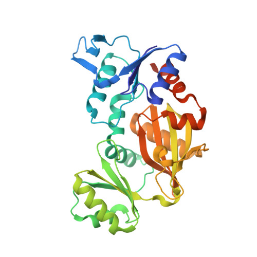
    Bacteria are equipped with a diverse set of regulatory tools that allow them to quickly adapt to their environment. The RimK system allows for Pseudomonas spp. to adapt through post-transcriptional regulation by altering the ribosomal subunit RpsF. RimK is found in a wide range of bacteria with a conserved amino acid sequence, however, the genetic context and the role of this protein is highly diverse. By solving and comparing the structures of RimK homologs from two related but functionally divergent systems, we uncovered key structural differences that likely contribute to the different activity levels of each of these homologs. Moreover, we were able to clearly resolve the active site of this protein for the first time, resolving binding of the glutamate substrate. This work advances our understanding of how subtle differences in protein sequence and structure can have profound effects on protein activity, which can in turn result in widespread mechanistic changes.


  • Organizational Affiliation

    Department of Molecular Microbiology, John Innes Centre, Norwich Research Park, Norwich, United Kingdom.


Macromolecules
Find similar proteins by:  (by identity cutoff)  |  3D Structure
Entity ID: 1
MoleculeChains Sequence LengthOrganismDetailsImage
Probable alpha-L-glutamate ligase
A, B, C, D, E
A, B, C, D, E, F, G, H, I, J, K, L, M, N, O, P
309Pseudomonas syringae pv. tomato str. DC3000Mutation(s): 0 
Gene Names: rimKPSPTO_0234
EC: 6.3.2
UniProt
Find proteins for Q88AZ9 (Pseudomonas syringae pv. tomato (strain ATCC BAA-871 / DC3000))
Explore Q88AZ9 
Go to UniProtKB:  Q88AZ9
Entity Groups  
Sequence Clusters30% Identity50% Identity70% Identity90% Identity95% Identity100% Identity
UniProt GroupQ88AZ9
Sequence Annotations
Expand
  • Reference Sequence
Small Molecules
Ligands 1 Unique
IDChains Name / Formula / InChI Key2D Diagram3D Interactions
ADP (Subject of Investigation/LOI)
Query on ADP

Download Ideal Coordinates CCD File 
AA [auth O]
Q [auth A]
R [auth B]
S [auth C]
T [auth D]
AA [auth O],
Q [auth A],
R [auth B],
S [auth C],
T [auth D],
U [auth E],
V [auth G],
W [auth H],
X [auth J],
Y [auth K],
Z [auth M]
ADENOSINE-5'-DIPHOSPHATE
C10 H15 N5 O10 P2
XTWYTFMLZFPYCI-KQYNXXCUSA-N
Experimental Data & Validation

Experimental Data

  • Method: X-RAY DIFFRACTION
  • Resolution: 2.90 Å
  • R-Value Free: 0.248 
  • R-Value Work: 0.237 
  • R-Value Observed: 0.238 
  • Space Group: P 1
Unit Cell:
Length ( Å )Angle ( ˚ )
a = 95.905α = 88.94
b = 96.091β = 84.37
c = 156.992γ = 89.97
Software Package:
Software NamePurpose
Aimlessdata scaling
REFMACrefinement
PDB_EXTRACTdata extraction
DIALSdata reduction
PHASERphasing

Structure Validation

View Full Validation Report


Ligand Structure Quality Assessment 


Entry History & Funding Information

Deposition Data


Funding OrganizationLocationGrant Number
Biotechnology and Biological Sciences Research Council (BBSRC)United KingdomBB/R018154/1
Biotechnology and Biological Sciences Research Council (BBSRC)United KingdomBBS/E/J/000PR9797
Biotechnology and Biological Sciences Research Council (BBSRC)United KingdomBBS/E/J/000PR9790

Revision History  (Full details and data files)

  • Version 1.0: 2022-10-05
    Type: Initial release
  • Version 1.1: 2023-02-08
    Changes: Database references
  • Version 1.2: 2024-02-07
    Changes: Data collection, Refinement description