7QNO

Crystal structure of ligand-free Danio rerio HDAC6 CD1 CD2


Experimental Data Snapshot

  • Method: X-RAY DIFFRACTION
  • Resolution: 2.38 Å
  • R-Value Free: 0.228 
  • R-Value Work: 0.212 
  • R-Value Observed: 0.213 

wwPDB Validation   3D Report Full Report


This is version 1.2 of the entry. See complete history


Literature

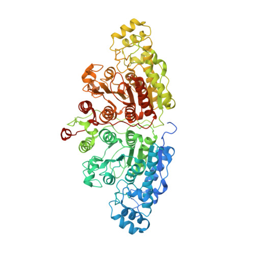
Expression and Crystallization of HDAC6 Tandem Catalytic Domains.

Langousis, G.Sanchez, J.Kempf, G.Matthias, P.

(2023) Methods Mol Biol 2589: 467-480

  • DOI: https://doi.org/10.1007/978-1-0716-2788-4_30
  • Primary Citation of Related Structures:  
    7QNO

  • PubMed Abstract: 

    Histone deacetylase 6 (HDAC6) is an atypical lysine deacetylase with tandem catalytic domains and an ubiquitin-binding zinc finger domain. HDAC6 is involved in various biological processes, such as cell motility or stress responses, and has been implicated in pathologies ranging from cancer to neurodegeneration. Due to this broad range of functions, there has been considerable interest in developing HDAC6-specific small molecule inhibitors, several of which are already available. The crystal structure of the tandem catalytic domains of zebrafish HDAC6 has revealed an arrangement with twofold symmetry and extensive surface interaction between the catalytic domains. Further dissection of the biochemical properties of HDAC6 and the development of novel inhibitors will benefit from being able to routinely express high-quality protein. We present here our optimized protocol for expression and crystallization of the zebrafish tandem catalytic domains.


  • Organizational Affiliation

    Friedrich Miescher Institute for Biomedical Research, Basel, Switzerland.


Macromolecules
Find similar proteins by:  (by identity cutoff)  |  3D Structure
Entity ID: 1
MoleculeChains Sequence LengthOrganismDetailsImage
Histone deacetylase 6837Danio rerioMutation(s): 0 
Gene Names: hdac6
UniProt
Find proteins for F8W4B7 (Danio rerio)
Explore F8W4B7 
Go to UniProtKB:  F8W4B7
Entity Groups  
Sequence Clusters30% Identity50% Identity70% Identity90% Identity95% Identity100% Identity
UniProt GroupF8W4B7
Sequence Annotations
Expand
  • Reference Sequence
Small Molecules
Ligands 4 Unique
IDChains Name / Formula / InChI Key2D Diagram3D Interactions
PRO
Query on PRO

Download Ideal Coordinates CCD File 
B [auth A]PROLINE
C5 H9 N O2
ONIBWKKTOPOVIA-BYPYZUCNSA-N
GOL
Query on GOL

Download Ideal Coordinates CCD File 
H [auth A]
I [auth A]
J [auth A]
K [auth A]
L [auth A]
H [auth A],
I [auth A],
J [auth A],
K [auth A],
L [auth A],
M [auth A]
GLYCEROL
C3 H8 O3
PEDCQBHIVMGVHV-UHFFFAOYSA-N
ZN
Query on ZN

Download Ideal Coordinates CCD File 
E [auth A]ZINC ION
Zn
PTFCDOFLOPIGGS-UHFFFAOYSA-N
K
Query on K

Download Ideal Coordinates CCD File 
C [auth A],
D [auth A],
F [auth A],
G [auth A]
POTASSIUM ION
K
NPYPAHLBTDXSSS-UHFFFAOYSA-N
Experimental Data & Validation

Experimental Data

  • Method: X-RAY DIFFRACTION
  • Resolution: 2.38 Å
  • R-Value Free: 0.228 
  • R-Value Work: 0.212 
  • R-Value Observed: 0.213 
  • Space Group: P 32 2 1
Unit Cell:
Length ( Å )Angle ( ˚ )
a = 186.134α = 90
b = 186.134β = 90
c = 102.442γ = 120
Software Package:
Software NamePurpose
PHENIXrefinement
Cootmodel building
autoPROCdata reduction
STARANISOdata scaling
PHASERphasing
ISOLDErefinement
XDSdata reduction

Structure Validation

View Full Validation Report


Entry History & Funding Information

Deposition Data


Funding OrganizationLocationGrant Number
Other private--

Revision History  (Full details and data files)

  • Version 1.0: 2022-02-09
    Type: Initial release
  • Version 1.1: 2022-11-23
    Changes: Database references, Refinement description
  • Version 1.2: 2024-01-31
    Changes: Data collection, Refinement description