7QGI

Crystal structure of SARS-CoV-2 NSP14 in the absence of NSP10


Experimental Data Snapshot

  • Method: X-RAY DIFFRACTION
  • Resolution: 1.65 Å
  • R-Value Free: 0.221 
  • R-Value Work: 0.196 
  • R-Value Observed: 0.197 

wwPDB Validation   3D Report Full Report


This is version 2.2 of the entry. See complete history


Literature

Crystal structures and fragment screening of SARS-CoV-2 NSP14 reveal details of exoribonuclease activation and mRNA capping and provide starting points for antiviral drug development.

Imprachim, N.Yosaatmadja, Y.Newman, J.A.

(2023) Nucleic Acids Res 51: 475-487

  • DOI: https://doi.org/10.1093/nar/gkac1207
  • Primary Citation of Related Structures:  
    7QGI, 7QIF

  • PubMed Abstract: 

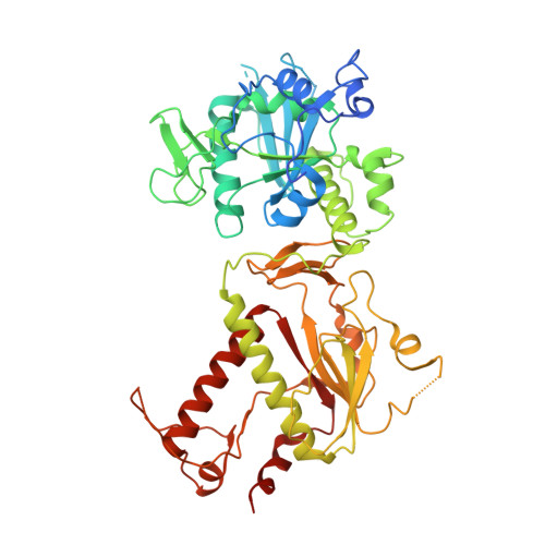
    NSP14 is a dual function enzyme containing an N-terminal exonuclease domain (ExoN) and C-terminal Guanine-N7-methyltransferase (N7-MTase) domain. Both activities are essential for the viral life cycle and may be targeted for anti-viral therapeutics. NSP14 forms a complex with NSP10, and this interaction enhances the nuclease but not the methyltransferase activity. We have determined the structure of SARS-CoV-2 NSP14 in the absence of NSP10 to 1.7 Å resolution. Comparisons with NSP14/NSP10 complexes reveal significant conformational changes that occur within the NSP14 ExoN domain upon binding of NSP10, including helix to coil transitions that facilitate the formation of the ExoN active site and provide an explanation of the stimulation of nuclease activity by NSP10. We have determined the structure of NSP14 in complex with cap analogue 7MeGpppG, and observe conformational changes within a SAM/SAH interacting loop that plays a key role in viral mRNA capping offering new insights into MTase activity. We perform an X-ray fragment screen on NSP14, revealing 72 hits bound to sites of inhibition in the ExoN and MTase domains. These fragments serve as excellent starting point tools for structure guided development of NSP14 inhibitors that may be used to treat COVID-19 and potentially other future viral threats.


  • Organizational Affiliation

    Centre for Medicines Discovery, University of Oxford, South Parks Rd, Oxford OX1 3QU, UK.


Macromolecules
Find similar proteins by:  (by identity cutoff)  |  3D Structure
Entity ID: 1
MoleculeChains Sequence LengthOrganismDetailsImage
Proofreading exoribonuclease nsp14A [auth D]523Severe acute respiratory syndrome coronavirus 2Mutation(s): 0 
Gene Names: rep1a-1b
EC: 3.1.13
UniProt
Find proteins for P0DTD1 (Severe acute respiratory syndrome coronavirus 2)
Explore P0DTD1 
Go to UniProtKB:  P0DTD1
Entity Groups  
Sequence Clusters30% Identity50% Identity70% Identity90% Identity95% Identity100% Identity
UniProt GroupP0DTD1
Sequence Annotations
Expand
  • Reference Sequence
Experimental Data & Validation

Experimental Data

  • Method: X-RAY DIFFRACTION
  • Resolution: 1.65 Å
  • R-Value Free: 0.221 
  • R-Value Work: 0.196 
  • R-Value Observed: 0.197 
  • Space Group: P 21 21 21
Unit Cell:
Length ( Å )Angle ( ˚ )
a = 67.539α = 90
b = 68.093β = 90
c = 138.428γ = 90
Software Package:
Software NamePurpose
PHENIXrefinement
PHENIXrefinement
XDSdata reduction
autoPROCdata scaling
PHASERphasing

Structure Validation

View Full Validation Report


Entry History & Funding Information

Deposition Data


Funding OrganizationLocationGrant Number
Not funded--

Revision History  (Full details and data files)

  • Version 1.0: 2022-01-26
    Type: Initial release
  • Version 2.0: 2022-07-20
    Type: Coordinate replacement
    Reason: Polymer geometry
    Changes: Advisory, Atomic model, Data collection, Database references, Derived calculations, Refinement description, Structure summary
  • Version 2.1: 2023-02-08
    Changes: Database references
  • Version 2.2: 2024-02-07
    Changes: Data collection, Refinement description