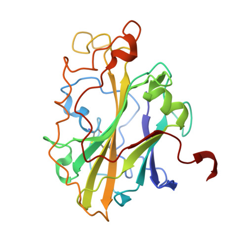
Protonation State of an Important Histidine from High Resolution Structures of Lytic Polysaccharide Monooxygenases.
Banerjee, S., Muderspach, S.J., Tandrup, T., Frandsen, K.E.H., Singh, R.K., Ipsen, J.O., Hernandez-Rollan, C., Norholm, M.H.H., Bjerrum, M.J., Johansen, K.S., Lo Leggio, L.(2022) Biomolecules 12
- PubMed: 35204695
- DOI: https://doi.org/10.3390/biom12020194
- Primary Citation of Related Structures:
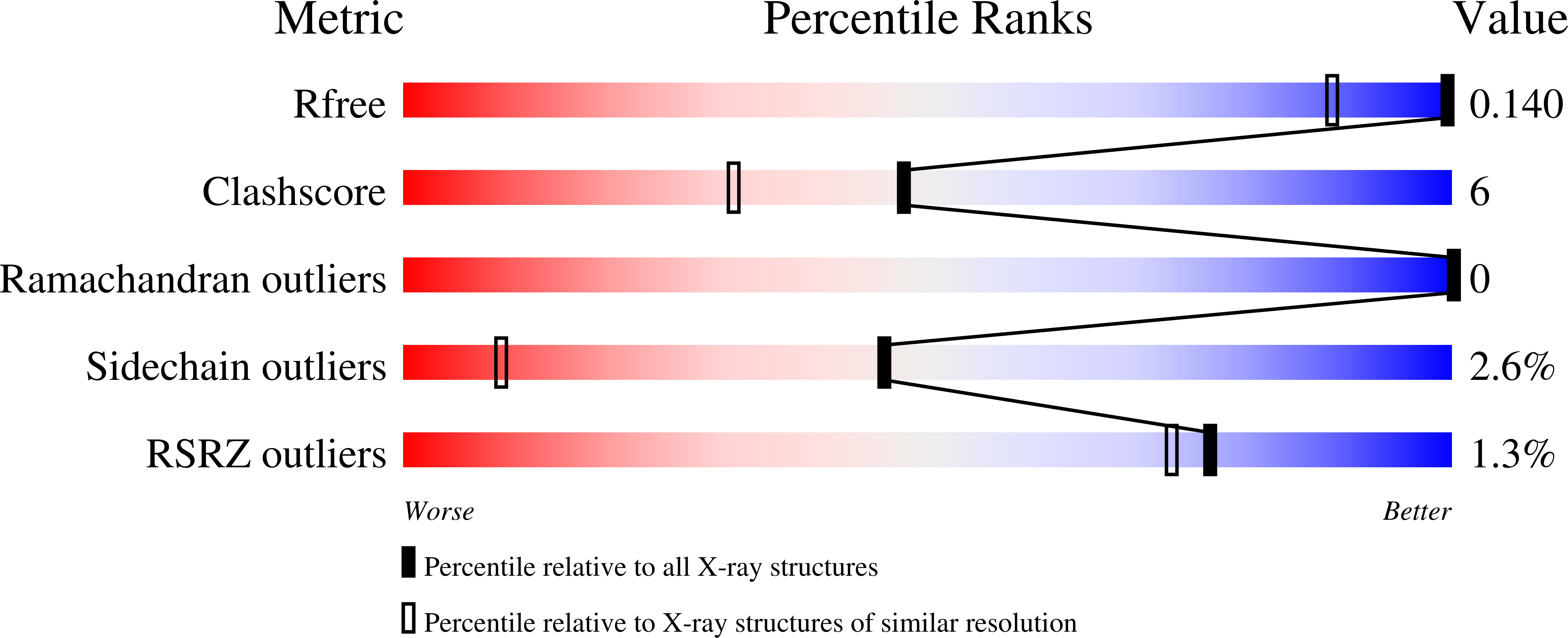
7PTZ, 7PU1 - PubMed Abstract:
Lytic Polysaccharide Monooxygenases (LPMOs) oxidatively cleave recalcitrant polysaccharides. The mechanism involves (i) reduction of the Cu, (ii) polysaccharide binding, (iii) binding of different oxygen species, and (iv) glycosidic bond cleavage. However, the complete mechanism is poorly understood and may vary across different families and even within the same family. Here, we have investigated the protonation state of a secondary co-ordination sphere histidine, conserved across AA9 family LPMOs that has previously been proposed to be a potential proton donor. Partial unrestrained refinement of newly obtained higher resolution data for two AA9 LPMOs and re-refinement of four additional data sets deposited in the PDB were carried out, where the His was refined without restraints, followed by measurements of the His ring geometrical parameters. This allowed reliable assignment of the protonation state, as also validated by following the same procedure for the His brace, for which the protonation state is predictable. The study shows that this histidine is generally singly protonated at the Nε2 atom, which is close to the oxygen species binding site. Our results indicate robustness of the method. In view of this and other emerging evidence, a role as proton donor during catalysis is unlikely for this His.
Organizational Affiliation:
Department of Chemistry, University of Copenhagen, Universitetsparken 5, DK-2100 Copenhagen, Denmark.