


Crystal structure of the allergen Jun a 3 the Thaumatin-like protein of Juniperus ashei
Eder, M., Hofer, G., Keller, W.To be published.
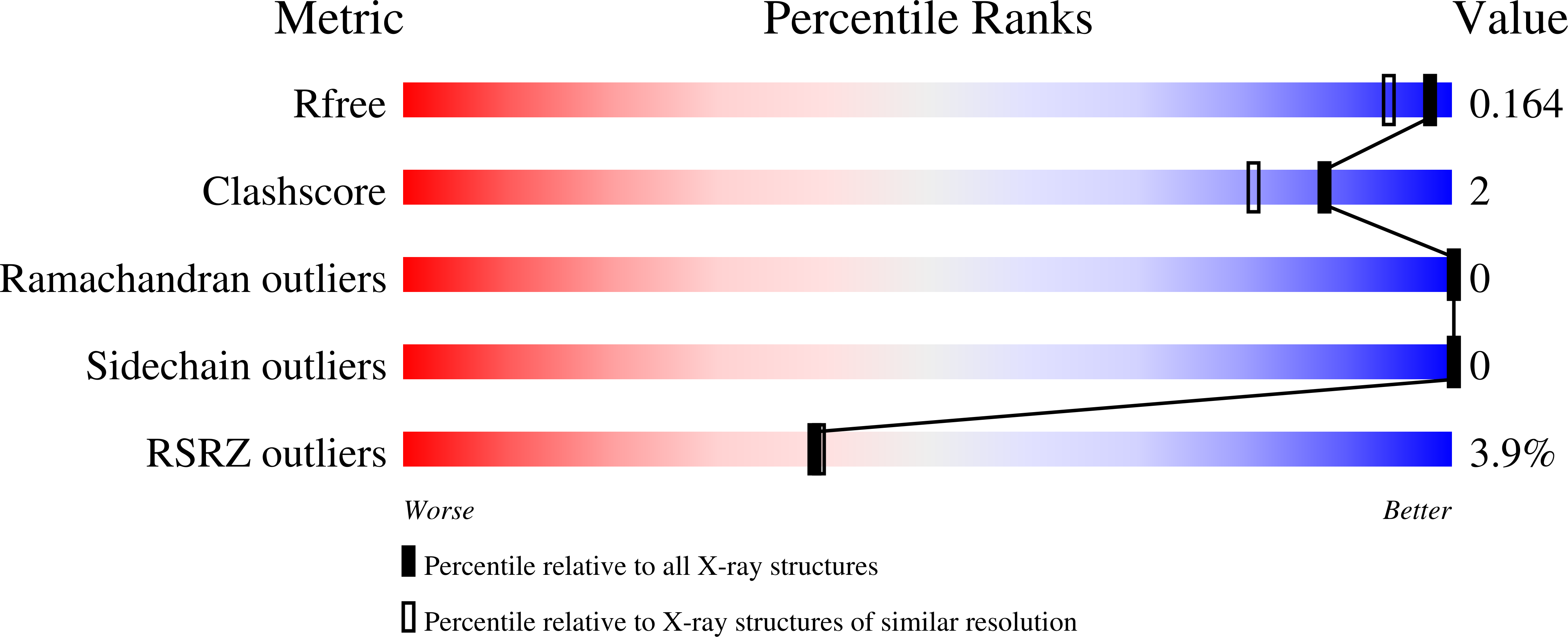
Experimental Data Snapshot
wwPDB Validation 3D Report Full Report
Entity ID: 1 | |||||
|---|---|---|---|---|---|
| Molecule | Chains | Sequence Length | Organism | Details | Image |
| Pathogenesis-related 5 protein Jun a 3.0101 | 207 | Juniperus ashei | Mutation(s): 2 | 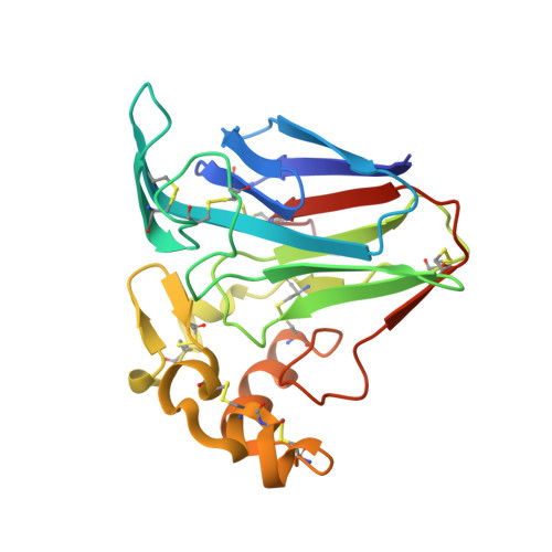 | |
UniProt | |||||
Find proteins for P81295 (Juniperus ashei) Explore P81295 Go to UniProtKB: P81295 | |||||
Entity Groups | |||||
| Sequence Clusters | 30% Identity50% Identity70% Identity90% Identity95% Identity100% Identity | ||||
| UniProt Group | P81295 | ||||
Sequence AnnotationsExpand | |||||
| |||||
| Ligands 1 Unique | |||||
|---|---|---|---|---|---|
| ID | Chains | Name / Formula / InChI Key | 2D Diagram | 3D Interactions | |
| CL Query on CL | B [auth A], C [auth A] | CHLORIDE ION Cl VEXZGXHMUGYJMC-UHFFFAOYSA-M |  | ||
| Length ( Å ) | Angle ( ˚ ) |
|---|---|
| a = 52.424 | α = 90 |
| b = 49.317 | β = 104.89 |
| c = 71.447 | γ = 90 |
| Software Name | Purpose |
|---|---|
| PHENIX | refinement |
| PDB_EXTRACT | data extraction |
| XDS | data reduction |
| XSCALE | data scaling |
| PHENIX | phasing |
| Funding Organization | Location | Grant Number |
|---|---|---|
| Austrian Science Fund | Austria | F 4604-B19 |
RCSB PDB (citation) is hosted by
RCSB PDB is a member of the

