7ODX

Cyanophage S-2L Succinoaminodeoxyadenylate synthetase (PurZ) bound to dGMP and dATP as an energy donor


Experimental Data Snapshot

  • Method: X-RAY DIFFRACTION
  • Resolution: 1.70 Å
  • R-Value Free: 0.176 
  • R-Value Work: 0.159 
  • R-Value Observed: 0.160 

wwPDB Validation   3D Report Full Report


Ligand Structure Quality Assessment 


This is version 1.1 of the entry. See complete history


Literature

Characterization of a triad of genes in cyanophage S-2L sufficient to replace adenine by 2-aminoadenine in bacterial DNA.

Czernecki, D.Bonhomme, F.Kaminski, P.A.Delarue, M.

(2021) Nat Commun 12: 4710-4710

  • DOI: https://doi.org/10.1038/s41467-021-25064-x
  • Primary Citation of Related Structures:  
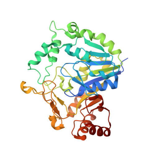
    7ODX, 7ODY

  • PubMed Abstract: 

    Cyanophage S-2L is known to profoundly alter the biophysical properties of its DNA by replacing all adenines (A) with 2-aminoadenines (Z), which still pair with thymines but with a triple hydrogen bond. It was recently demonstrated that a homologue of adenylosuccinate synthetase (PurZ) and a dATP triphosphohydrolase (DatZ) are two important pieces of the metabolism of 2-aminoadenine, participating in the synthesis of ZTGC-DNA. Here, we determine that S-2L PurZ can use either dATP or ATP as a source of energy, thereby also depleting the pool of nucleotides in dATP. Furthermore, we identify a conserved gene (mazZ) located between purZ and datZ genes in S-2L and related phage genomes. We show that it encodes a (d)GTP-specific diphosphohydrolase, thereby providing the substrate of PurZ in the 2-aminoadenine synthesis pathway. High-resolution crystal structures of S-2L PurZ and MazZ with their respective substrates provide a rationale for their specificities. The Z-cluster made of these three genes - datZ, mazZ and purZ - was expressed in E. coli, resulting in a successful incorporation of 2-aminoadenine in the bacterial chromosomal and plasmidic DNA. This work opens the possibility to study synthetic organisms containing ZTGC-DNA.


  • Organizational Affiliation

    Unit of Architecture and Dynamics of Biological Macromolecules, CNRS UMR 3528, 25-28 rue du Docteur Roux, Institut Pasteur, Paris, France.


Macromolecules
Find similar proteins by:  (by identity cutoff)  |  3D Structure
Entity ID: 1
MoleculeChains Sequence LengthOrganismDetailsImage
Succinoaminodeoxyadenylate synthetase (PurZ)365Cyanophage S-2LMutation(s): 0 
UniProt
Find proteins for A0A7U3TBV6 (Cyanophage S-2L)
Explore A0A7U3TBV6 
Go to UniProtKB:  A0A7U3TBV6
Entity Groups  
Sequence Clusters30% Identity50% Identity70% Identity90% Identity95% Identity100% Identity
UniProt GroupA0A7U3TBV6
Sequence Annotations
Expand
  • Reference Sequence
Small Molecules
Ligands 2 Unique
IDChains Name / Formula / InChI Key2D Diagram3D Interactions
DTP (Subject of Investigation/LOI)
Query on DTP

Download Ideal Coordinates CCD File 
C [auth A]2'-DEOXYADENOSINE 5'-TRIPHOSPHATE
C10 H16 N5 O12 P3
SUYVUBYJARFZHO-RRKCRQDMSA-N
DGP (Subject of Investigation/LOI)
Query on DGP

Download Ideal Coordinates CCD File 
B [auth A]2'-DEOXYGUANOSINE-5'-MONOPHOSPHATE
C10 H14 N5 O7 P
LTFMZDNNPPEQNG-KVQBGUIXSA-N
Experimental Data & Validation

Experimental Data

  • Method: X-RAY DIFFRACTION
  • Resolution: 1.70 Å
  • R-Value Free: 0.176 
  • R-Value Work: 0.159 
  • R-Value Observed: 0.160 
  • Space Group: P 62 2 2
Unit Cell:
Length ( Å )Angle ( ˚ )
a = 108.18α = 90
b = 108.18β = 90
c = 142.33γ = 120
Software Package:
Software NamePurpose
PHENIXrefinement
XDSdata reduction
XDSdata scaling
PHASERphasing

Structure Validation

View Full Validation Report


Ligand Structure Quality Assessment 


Entry History 

Deposition Data

Revision History  (Full details and data files)

  • Version 1.0: 2021-09-01
    Type: Initial release
  • Version 1.1: 2024-01-31
    Changes: Data collection, Refinement description