7NEF

Fucosylated linear peptide Fln65 bound to the fucose binding lectin LecB PA-IIL from Pseudomonas aeruginosa at 1.5 Angstrom resolution


Experimental Data Snapshot

  • Method: X-RAY DIFFRACTION
  • Resolution: 1.51 Å
  • R-Value Free: 0.206 
  • R-Value Work: 0.182 
  • R-Value Observed: 0.183 

wwPDB Validation   3D Report Full Report


Ligand Structure Quality Assessment 


This is version 1.2 of the entry. See complete history


Literature

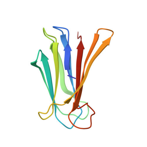
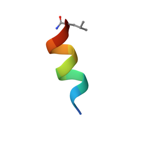
A mixed chirality alpha-helix in a stapled bicyclic and a linear antimicrobial peptide revealed by X-ray crystallography.

Baeriswyl, S.Personne, H.Di Bonaventura, I.Kohler, T.van Delden, C.Stocker, A.Javor, S.Reymond, J.L.

(2021) RSC Chem Biol 2: 1608-1617

  • DOI: https://doi.org/10.1039/d1cb00124h
  • Primary Citation of Related Structures:  
    6Y0U, 6Y0V, 6Y13, 6Y14, 6Y1S, 7NEF, 7NEW

  • PubMed Abstract: 

    The peptide α-helix is right-handed when containing amino acids with l-chirality, and left-handed with d-chirality, however mixed chirality peptides generally do not form α-helices unless a helix inducer such as the non-natural residue amino-isobutyric acid is used. Herein we report the first X-ray crystal structures of mixed chirality α-helices in short peptides comprising only natural residues as the example of a stapled bicyclic and a linear membrane disruptive amphiphilic antimicrobial peptide (AMP) containing seven l- and four d-residues, as complexes of fucosylated analogs with the bacterial lectin LecB. The mixed chirality α-helices are superimposable onto the homochiral α-helices and form under similar conditions as shown by CD spectra and MD simulations but non-hemolytic and resistant to proteolysis. The observation of a mixed chirality α-helix with only natural residues in the protein environment of LecB suggests a vast unexplored territory of α-helical mixed chirality sequences and their possible use for optimizing bioactive α-helical peptides.


  • Organizational Affiliation

    Department of Chemistry, Biochemistry and Pharmaceutical Sciences, University of Bern Freiestrasse 3 3012 Bern Switzerland jean-louis.reymond@unibe.ch.


Macromolecules
Find similar proteins by:  (by identity cutoff)  |  3D Structure
Entity ID: 1
MoleculeChains Sequence LengthOrganismDetailsImage
Fucose-binding lectin
A, B, C, D, E
A, B, C, D, E, F, G, H
115Pseudomonas aeruginosaMutation(s): 0 
Gene Names: 
UniProt
Find proteins for Q9HYN5 (Pseudomonas aeruginosa (strain ATCC 15692 / DSM 22644 / CIP 104116 / JCM 14847 / LMG 12228 / 1C / PRS 101 / PAO1))
Explore Q9HYN5 
Go to UniProtKB:  Q9HYN5
Entity Groups  
Sequence Clusters30% Identity50% Identity70% Identity90% Identity95% Identity100% Identity
UniProt GroupQ9HYN5
Sequence Annotations
Expand
  • Reference Sequence

Find similar proteins by:  Sequence   |   3D Structure  

Entity ID: 2
MoleculeChains Sequence LengthOrganismDetailsImage
Fln65
I, J, K, L, M
I, J, K, L, M, N, O, P
12synthetic constructMutation(s): 0 
Entity Groups  
Sequence Clusters30% Identity50% Identity70% Identity90% Identity95% Identity100% Identity
Sequence Annotations
Expand
  • Reference Sequence
Small Molecules
Ligands 2 Unique
IDChains Name / Formula / InChI Key2D Diagram3D Interactions
ZDC
Query on ZDC

Download Ideal Coordinates CCD File 
GA [auth I]
HA [auth J]
IA [auth K]
JA [auth L]
KA [auth M]
GA [auth I],
HA [auth J],
IA [auth K],
JA [auth L],
KA [auth M],
LA [auth N],
MA [auth O],
NA [auth P]
3,7-anhydro-2,8-dideoxy-L-glycero-D-gluco-octonic acid
C8 H14 O6
YTZUDUWVQZSKNN-OASCRQMUSA-N
CA
Query on CA

Download Ideal Coordinates CCD File 
AA [auth F]
BA [auth F]
CA [auth G]
DA [auth G]
EA [auth H]
AA [auth F],
BA [auth F],
CA [auth G],
DA [auth G],
EA [auth H],
FA [auth H],
Q [auth A],
R [auth A],
S [auth B],
T [auth B],
U [auth C],
V [auth C],
W [auth D],
X [auth D],
Y [auth E],
Z [auth E]
CALCIUM ION
Ca
BHPQYMZQTOCNFJ-UHFFFAOYSA-N
Experimental Data & Validation

Experimental Data

  • Method: X-RAY DIFFRACTION
  • Resolution: 1.51 Å
  • R-Value Free: 0.206 
  • R-Value Work: 0.182 
  • R-Value Observed: 0.183 
  • Space Group: P 1 21 1
Unit Cell:
Length ( Å )Angle ( ˚ )
a = 74.467α = 90
b = 64.3β = 94.91
c = 118.926γ = 90
Software Package:
Software NamePurpose
XDSdata reduction
XSCALEdata scaling
PHASERphasing
PHENIXrefinement
PDB_EXTRACTdata extraction

Structure Validation

View Full Validation Report


Ligand Structure Quality Assessment 


Entry History & Funding Information

Deposition Data


Funding OrganizationLocationGrant Number
Swiss National Science FoundationSwitzerland--

Revision History  (Full details and data files)

  • Version 1.0: 2021-03-03
    Type: Initial release
  • Version 1.1: 2022-02-02
    Changes: Database references
  • Version 1.2: 2024-01-31
    Changes: Data collection, Database references, Refinement description