7N89

Room-temperature X-ray structure of SARS-CoV-2 main protease C145A mutant in complex with substrate Ac-SAVLQSGF-CONH2


Experimental Data Snapshot

  • Method: X-RAY DIFFRACTION
  • Resolution: 2.00 Å
  • R-Value Free: 0.218 
  • R-Value Work: 0.171 
  • R-Value Observed: 0.173 

wwPDB Validation   3D Report Full Report


This is version 1.2 of the entry. See complete history


Literature

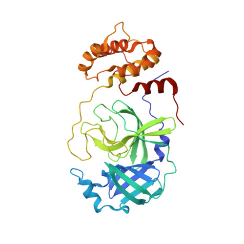
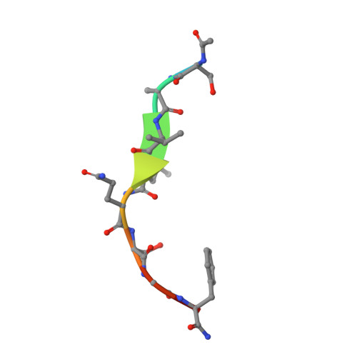
Michaelis-like complex of SARS-CoV-2 main protease visualized by room-temperature X-ray crystallography.

Kneller, D.W.Zhang, Q.Coates, L.Louis, J.M.Kovalevsky, A.

(2021) IUCrJ 8: 973-979

  • DOI: https://doi.org/10.1107/S2052252521010113
  • Primary Citation of Related Structures:  
    7N89

  • PubMed Abstract: 

    SARS-CoV-2 emerged at the end of 2019 to cause an unprecedented pandemic of the deadly respiratory disease COVID-19 that continues to date. The viral main protease (M pro ) is essential for SARS-CoV-2 replication and is therefore an important drug target. Understanding the catalytic mechanism of M pro , a cysteine protease with a catalytic site comprising the noncanonical Cys145-His41 dyad, can help in guiding drug design. Here, a 2.0 Å resolution room-temperature X-ray crystal structure is reported of a Michaelis-like complex of M pro harboring a single inactivating mutation C145A bound to the octapeptide Ac-SAVLQSGF-CONH 2 corresponding to the nsp4/nsp5 autocleavage site. The peptide substrate is unambiguously defined in subsites S5 to S3' by strong electron density. Superposition of the Michaelis-like complex with the neutron structure of substrate-free M pro demonstrates that the catalytic site is inherently pre-organized for catalysis prior to substrate binding. Induced fit to the substrate is driven by P1 Gln binding in the predetermined subsite S1 and rearrangement of subsite S2 to accommodate P2 Leu. The Michaelis-like complex structure is ideal for in silico modeling of the SARS-CoV-2 M pro catalytic mechanism.


  • Organizational Affiliation

    Neutron Scattering Division, Oak Ridge National Laboratory, 1 Bethel Valley Road, Oak Ridge, TN 37831, USA.


Macromolecules
Find similar proteins by:  (by identity cutoff)  |  3D Structure
Entity ID: 1
MoleculeChains Sequence LengthOrganismDetailsImage
3C-like proteinase
A, B
306Severe acute respiratory syndrome coronavirus 2Mutation(s): 1 
Gene Names: rep1a-1b
EC: 3.4.22.69
UniProt
Find proteins for P0DTD1 (Severe acute respiratory syndrome coronavirus 2)
Explore P0DTD1 
Go to UniProtKB:  P0DTD1
Entity Groups  
Sequence Clusters30% Identity50% Identity70% Identity90% Identity95% Identity100% Identity
UniProt GroupP0DTD1
Sequence Annotations
Expand
  • Reference Sequence

Find similar proteins by:  Sequence   |   3D Structure  

Entity ID: 2
MoleculeChains Sequence LengthOrganismDetailsImage
ACE-SER-ALA-VAL-LEU-GLN-SER-GLY-PHE-NH2
C, D
10Severe acute respiratory syndrome coronavirus 2Mutation(s): 0 
UniProt
Find proteins for P0DTD1 (Severe acute respiratory syndrome coronavirus 2)
Explore P0DTD1 
Go to UniProtKB:  P0DTD1
Entity Groups  
Sequence Clusters30% Identity50% Identity70% Identity90% Identity95% Identity100% Identity
UniProt GroupP0DTD1
Sequence Annotations
Expand
  • Reference Sequence
Experimental Data & Validation

Experimental Data

  • Method: X-RAY DIFFRACTION
  • Resolution: 2.00 Å
  • R-Value Free: 0.218 
  • R-Value Work: 0.171 
  • R-Value Observed: 0.173 
  • Space Group: P 1
Unit Cell:
Length ( Å )Angle ( ˚ )
a = 47.654α = 66.88
b = 54.104β = 79.02
c = 60.496γ = 88.52
Software Package:
Software NamePurpose
PHENIXrefinement
CrysalisProdata reduction
Aimlessdata scaling
PHASERphasing

Structure Validation

View Full Validation Report


Entry History 

Deposition Data

Revision History  (Full details and data files)

  • Version 1.0: 2021-06-23
    Type: Initial release
  • Version 1.1: 2021-12-15
    Changes: Database references
  • Version 1.2: 2023-10-18
    Changes: Data collection, Refinement description