7MMD

Crystal structure of HCV NS3/4A D168A protease in complex with JZ01-19

  • Classification: HYDROLASE/INHIBITOR
  • Organism(s): Hepacivirus hominis
  • Expression System: Escherichia coli BL21(DE3)
  • Mutation(s): Yes 

  • Deposited: 2021-04-29 Released: 2022-08-31 
  • Deposition Author(s): Zephyr, J., Schiffer, C.A.
  • Funding Organization(s): National Institutes of Health/National Institute Of Allergy and Infectious Diseases (NIH/NIAID), National Institutes of Health/National Institute of General Medical Sciences (NIH/NIGMS)

Experimental Data Snapshot

  • Method: X-RAY DIFFRACTION
  • Resolution: 1.89 Å
  • R-Value Free: 0.198 
  • R-Value Work: 0.160 
  • R-Value Observed: 0.161 

wwPDB Validation   3D Report Full Report


Ligand Structure Quality Assessment 


This is version 1.1 of the entry. See complete history


Literature

Deciphering the Molecular Mechanism of HCV Protease Inhibitor Fluorination as a General Approach to Avoid Drug Resistance.

Zephyr, J.Nageswara Rao, D.Vo, S.V.Henes, M.Kosovrasti, K.Matthew, A.N.Hedger, A.K.Timm, J.Chan, E.T.Ali, A.Kurt Yilmaz, N.Schiffer, C.A.

(2022) J Mol Biol 434: 167503-167503

  • DOI: https://doi.org/10.1016/j.jmb.2022.167503
  • Primary Citation of Related Structures:  
    7MM2, 7MM3, 7MM4, 7MM5, 7MM6, 7MM7, 7MM8, 7MM9, 7MMA, 7MMB, 7MMC, 7MMD, 7MMF, 7MMG, 7MMH, 7MMI, 7MMJ, 7MMK, 7MML

  • PubMed Abstract: 

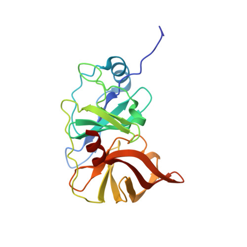
    Third generation Hepatitis C virus (HCV) NS3/4A protease inhibitors (PIs), glecaprevir and voxilaprevir, are highly effective across genotypes and against many resistant variants. Unlike earlier PIs, these compounds have fluorine substitutions on the P2-P4 macrocycle and P1 moieties. Fluorination has long been used in medicinal chemistry as a strategy to improve physicochemical properties and potency. However, the molecular basis by which fluorination improves potency and resistance profile of HCV NS3/4A PIs is not well understood. To systematically analyze the contribution of fluorine substitutions to inhibitor potency and resistance profile, we used a multi-disciplinary approach involving inhibitor design and synthesis, enzyme inhibition assays, co-crystallography, and structural analysis. A panel of inhibitors in matched pairs were designed with and without P4 cap fluorination, tested against WT protease and the D168A resistant variant, and a total of 22 high-resolution co-crystal structures were determined. While fluorination did not significantly improve potency against the WT protease, PIs with fluorinated P4 caps retained much better potency against the D168A protease variant. Detailed analysis of the co-crystal structures revealed that PIs with fluorinated P4 caps can sample alternate binding conformations that enable adapting to structural changes induced by the D168A substitution. Our results elucidate molecular mechanisms of fluorine-specific inhibitor interactions that can be leveraged in avoiding drug resistance.


  • Organizational Affiliation

    Department of Biochemistry and Molecular Pharmacology, University of Massachusetts Medical School, Worcester, MA 01605, United States.


Macromolecules
Find similar proteins by:  (by identity cutoff)  |  3D Structure
Entity ID: 1
MoleculeChains Sequence LengthOrganismDetailsImage
NS3/4A protease200Hepacivirus hominisMutation(s): 1 
UniProt
Find proteins for P26664 (Hepatitis C virus genotype 1a (isolate 1))
Explore P26664 
Go to UniProtKB:  P26664
Entity Groups  
Sequence Clusters30% Identity50% Identity70% Identity90% Identity95% Identity100% Identity
UniProt GroupP26664
Sequence Annotations
Expand
  • Reference Sequence
Small Molecules
Ligands 4 Unique
IDChains Name / Formula / InChI Key2D Diagram3D Interactions
ZJP (Subject of Investigation/LOI)
Query on ZJP

Download Ideal Coordinates CCD File 
G [auth A]cyclopentyl {(2R,4S,6S,12Z,13aS,14aR,16aS)-2-[(7-methoxy-3-methylquinoxalin-2-yl)oxy]-14a-[(1-methylcyclopropane-1-sulfonyl)carbamoyl]-5,16-dioxo-1,2,3,5,6,7,8,9,10,11,13a,14,14a,15,16,16a-hexadecahydrocyclopropa[e]pyrrolo[1,2-a][1,4]diazacyclopentadecin-6-yl}carbamate
C38 H50 N6 O9 S
AWCLTDQQVULZEM-UIWVBJMMSA-N
SO4
Query on SO4

Download Ideal Coordinates CCD File 
I [auth A]SULFATE ION
O4 S
QAOWNCQODCNURD-UHFFFAOYSA-L
ZN
Query on ZN

Download Ideal Coordinates CCD File 
D [auth A]ZINC ION
Zn
PTFCDOFLOPIGGS-UHFFFAOYSA-N
EDO
Query on EDO

Download Ideal Coordinates CCD File 
B [auth A],
C [auth A],
E [auth A],
F [auth A],
H [auth A]
1,2-ETHANEDIOL
C2 H6 O2
LYCAIKOWRPUZTN-UHFFFAOYSA-N
Experimental Data & Validation

Experimental Data

  • Method: X-RAY DIFFRACTION
  • Resolution: 1.89 Å
  • R-Value Free: 0.198 
  • R-Value Work: 0.160 
  • R-Value Observed: 0.161 
  • Space Group: P 21 21 21
Unit Cell:
Length ( Å )Angle ( ˚ )
a = 55.151α = 90
b = 58.7β = 90
c = 59.858γ = 90
Software Package:
Software NamePurpose
PHENIXrefinement
PHASERphasing
HKL-3000data scaling
Cootmodel building

Structure Validation

View Full Validation Report


Ligand Structure Quality Assessment 


Entry History & Funding Information

Deposition Data


Funding OrganizationLocationGrant Number
National Institutes of Health/National Institute Of Allergy and Infectious Diseases (NIH/NIAID)United StatesR01 AI085051
National Institutes of Health/National Institute of General Medical Sciences (NIH/NIGMS)United StatesF31 GM131635

Revision History  (Full details and data files)

  • Version 1.0: 2022-08-31
    Type: Initial release
  • Version 1.1: 2023-10-18
    Changes: Data collection, Refinement description