7MC6

Crystal structure of the SARS-CoV-2 ExoN-nsp10 complex containing Mg2+ ion


Experimental Data Snapshot

  • Method: X-RAY DIFFRACTION
  • Resolution: 2.10 Å
  • R-Value Free: 0.219 
  • R-Value Work: 0.197 
  • R-Value Observed: 0.198 

wwPDB Validation   3D Report Full Report


This is version 1.2 of the entry. See complete history


Literature

Structure and dynamics of SARS-CoV-2 proofreading exoribonuclease ExoN.

Moeller, N.H.Shi, K.Demir, O.Belica, C.Banerjee, S.Yin, L.Durfee, C.Amaro, R.E.Aihara, H.

(2022) Proc Natl Acad Sci U S A 119

  • DOI: https://doi.org/10.1073/pnas.2106379119
  • Primary Citation of Related Structures:  
    7MC5, 7MC6

  • PubMed Abstract: 

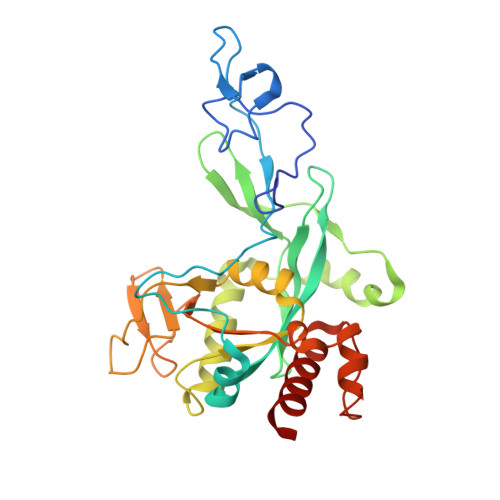
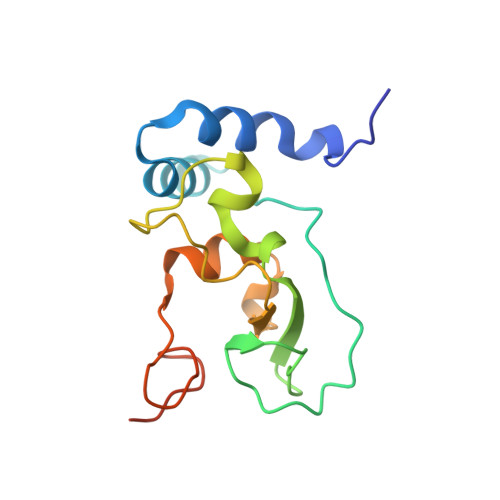
    High-fidelity replication of the large RNA genome of coronaviruses (CoVs) is mediated by a 3'-to-5' exoribonuclease (ExoN) in nonstructural protein 14 (nsp14), which excises nucleotides including antiviral drugs misincorporated by the low-fidelity viral RNA-dependent RNA polymerase (RdRp) and has also been implicated in viral RNA recombination and resistance to innate immunity. Here, we determined a 1.6-Å resolution crystal structure of severe acute respiratory syndrome CoV 2 (SARS-CoV-2) ExoN in complex with its essential cofactor, nsp10. The structure shows a highly basic and concave surface flanking the active site, comprising several Lys residues of nsp14 and the N-terminal amino group of nsp10. Modeling suggests that this basic patch binds to the template strand of double-stranded RNA substrates to position the 3' end of the nascent strand in the ExoN active site, which is corroborated by mutational and computational analyses. We also show that the ExoN activity can rescue a stalled RNA primer poisoned with sofosbuvir and allow RdRp to continue its extension in the presence of the chain-terminating drug, biochemically recapitulating proofreading in SARS-CoV-2 replication. Molecular dynamics simulations further show remarkable flexibility of multidomain nsp14 and suggest that nsp10 stabilizes ExoN for substrate RNA binding to support its exonuclease activity. Our high-resolution structure of the SARS-CoV-2 ExoN-nsp10 complex serves as a platform for future development of anticoronaviral drugs or strategies to attenuate the viral virulence.


  • Organizational Affiliation

    Department of Biochemistry, Molecular Biology and Biophysics, University of Minnesota, Minneapolis, MN 55455.


Macromolecules
Find similar proteins by:  (by identity cutoff)  |  3D Structure
Entity ID: 1
MoleculeChains Sequence LengthOrganismDetailsImage
Proofreading exoribonuclease291Severe acute respiratory syndrome coronavirus 2Mutation(s): 0 
Gene Names: rep1a-1b
EC: 3.1.13
UniProt
Find proteins for P0DTD1 (Severe acute respiratory syndrome coronavirus 2)
Explore P0DTD1 
Go to UniProtKB:  P0DTD1
Entity Groups  
Sequence Clusters30% Identity50% Identity70% Identity90% Identity95% Identity100% Identity
UniProt GroupP0DTD1
Sequence Annotations
Expand
  • Reference Sequence
Find similar proteins by:  (by identity cutoff)  |  3D Structure
Entity ID: 2
MoleculeChains Sequence LengthOrganismDetailsImage
Non-structural protein 10B [auth M]140Severe acute respiratory syndrome coronavirus 2Mutation(s): 0 
Gene Names: rep1a-1b
UniProt
Find proteins for P0DTD1 (Severe acute respiratory syndrome coronavirus 2)
Explore P0DTD1 
Go to UniProtKB:  P0DTD1
Entity Groups  
Sequence Clusters30% Identity50% Identity70% Identity90% Identity95% Identity100% Identity
UniProt GroupP0DTD1
Sequence Annotations
Expand
  • Reference Sequence
Small Molecules
Ligands 4 Unique
IDChains Name / Formula / InChI Key2D Diagram3D Interactions
ZN
Query on ZN

Download Ideal Coordinates CCD File 
C [auth A],
D [auth A],
N [auth M],
O [auth M]
ZINC ION
Zn
PTFCDOFLOPIGGS-UHFFFAOYSA-N
EDO
Query on EDO

Download Ideal Coordinates CCD File 
E [auth A]
F [auth A]
G [auth A]
H [auth A]
I [auth A]
E [auth A],
F [auth A],
G [auth A],
H [auth A],
I [auth A],
J [auth A],
K [auth A],
P [auth M],
Q [auth M]
1,2-ETHANEDIOL
C2 H6 O2
LYCAIKOWRPUZTN-UHFFFAOYSA-N
CL
Query on CL

Download Ideal Coordinates CCD File 
L [auth A]CHLORIDE ION
Cl
VEXZGXHMUGYJMC-UHFFFAOYSA-M
MG
Query on MG

Download Ideal Coordinates CCD File 
M [auth A]MAGNESIUM ION
Mg
JLVVSXFLKOJNIY-UHFFFAOYSA-N
Experimental Data & Validation

Experimental Data

  • Method: X-RAY DIFFRACTION
  • Resolution: 2.10 Å
  • R-Value Free: 0.219 
  • R-Value Work: 0.197 
  • R-Value Observed: 0.198 
  • Space Group: P 21 21 21
Unit Cell:
Length ( Å )Angle ( ˚ )
a = 61.67α = 90
b = 70.32β = 90
c = 108.54γ = 90
Software Package:
Software NamePurpose
Aimlessdata scaling
PHENIXrefinement
PDB_EXTRACTdata extraction
XDSdata reduction
PHASERphasing

Structure Validation

View Full Validation Report


Entry History & Funding Information

Deposition Data


Funding OrganizationLocationGrant Number
National Institutes of Health/National Institute of General Medical Sciences (NIH/NIGMS)United StatesGM118047

Revision History  (Full details and data files)

  • Version 1.0: 2021-05-05
    Type: Initial release
  • Version 1.1: 2022-03-16
    Changes: Database references
  • Version 1.2: 2023-10-18
    Changes: Data collection, Database references, Refinement description