Molecular Dynamics Free Energy Simulations Reveal the Mechanism for the Antiviral Resistance of the M66I HIV-1 Capsid Mutation.
Sun, Q., Levy, R.M., Kirby, K.A., Wang, Z., Sarafianos, S.G., Deng, N.(2021) Viruses 13
- PubMed: 34063519
- DOI: https://doi.org/10.3390/v13050920
- Primary Citation of Related Structures:
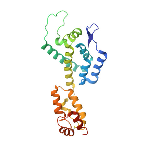
7M9F - PubMed Abstract:
While drug resistance mutations can often be attributed to the loss of direct or solvent-mediated protein-ligand interactions in the drug-mutant complex, in this study we show that a resistance mutation for the picomolar HIV-1 capsid (CA)-targeting antiviral (GS-6207) is mainly due to the free energy cost of the drug-induced protein side chain reorganization in the mutant protein. Among several mutations, M66I causes the most suppression of the GS-6207 antiviral activity (up to ~84,000-fold), and only 83- and 68-fold reductions for PF74 and ZW-1261, respectively. To understand the molecular basis of this drug resistance, we conducted molecular dynamics free energy simulations to study the structures, energetics, and conformational free energy landscapes involved in the inhibitors binding at the interface of two CA monomers. To minimize the protein-ligand steric clash, the I66 side chain in the M66I-GS-6207 complex switches to a higher free energy conformation from the one adopted in the apo M66I. In contrast, the binding of GS-6207 to the wild-type CA does not lead to any significant M66 conformational change. Based on an analysis that decomposes the absolute binding free energy into contributions from two receptor conformational states, it appears that it is the free energy cost of side chain reorganization rather than the reduced protein-ligand interaction that is largely responsible for the drug resistance against GS-6207.
Organizational Affiliation:
Center for Biophysics and Computational Biology and Department of Chemistry, Temple University, Philadelphia, PA 19122, USA.