7LUA

Cryo-EM structure of DH898.1 Fab-dimer bound near the CD4 binding site of HIV-1 Env CH848 SOSIP trimer


Experimental Data Snapshot

  • Method: ELECTRON MICROSCOPY
  • Resolution: 4.70 Å
  • Aggregation State: PARTICLE 
  • Reconstruction Method: SINGLE PARTICLE 

wwPDB Validation   3D Report Full Report


This is version 1.2 of the entry. See complete history


Literature

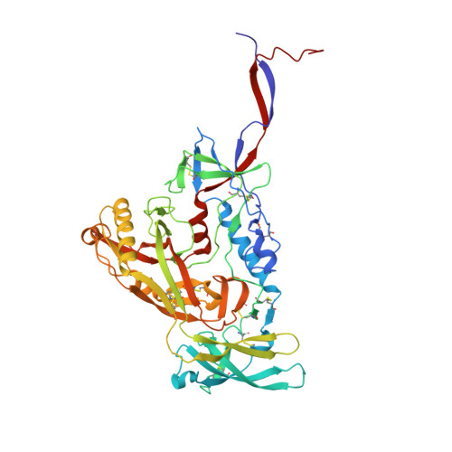
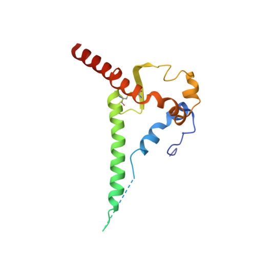
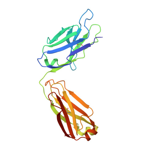
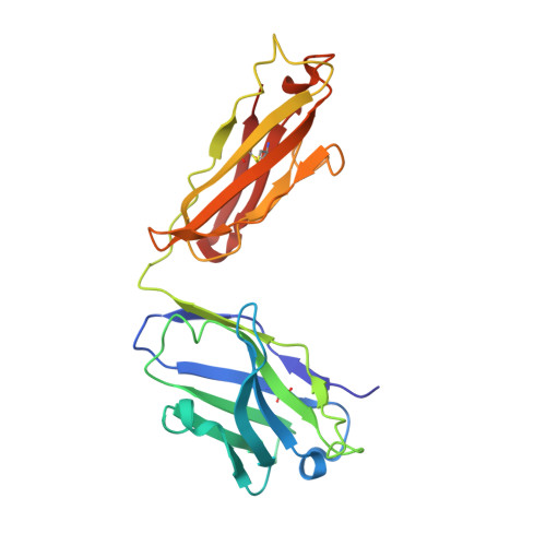
Fab-dimerized glycan-reactive antibodies are a structural category of natural antibodies.

Williams, W.B.Meyerhoff, R.R.Edwards, R.J.Li, H.Manne, K.Nicely, N.I.Henderson, R.Zhou, Y.Janowska, K.Mansouri, K.Gobeil, S.Evangelous, T.Hora, B.Berry, M.Abuahmad, A.Y.Sprenz, J.Deyton, M.Stalls, V.Kopp, M.Hsu, A.L.Borgnia, M.J.Stewart-Jones, G.B.E.Lee, M.S.Bronkema, N.Moody, M.A.Wiehe, K.Bradley, T.Alam, S.M.Parks, R.J.Foulger, A.Oguin, T.Sempowski, G.D.Bonsignori, M.LaBranche, C.C.Montefiori, D.C.Seaman, M.Santra, S.Perfect, J.Francica, J.R.Lynn, G.M.Aussedat, B.Walkowicz, W.E.Laga, R.Kelsoe, G.Saunders, K.O.Fera, D.Kwong, P.D.Seder, R.A.Bartesaghi, A.Shaw, G.M.Acharya, P.Haynes, B.F.

(2021) Cell 184: 2955

  • DOI: https://doi.org/10.1016/j.cell.2021.04.042
  • Primary Citation of Related Structures:  
    6VTU, 6XRJ, 7L02, 7L06, 7L09, 7L6M, 7L6O, 7LU9, 7LUA

  • PubMed Abstract: 

    Natural antibodies (Abs) can target host glycans on the surface of pathogens. We studied the evolution of glycan-reactive B cells of rhesus macaques and humans using glycosylated HIV-1 envelope (Env) as a model antigen. 2G12 is a broadly neutralizing Ab (bnAb) that targets a conserved glycan patch on Env of geographically diverse HIV-1 strains using a unique heavy-chain (V H ) domain-swapped architecture that results in fragment antigen-binding (Fab) dimerization. Here, we describe HIV-1 Env Fab-dimerized glycan (FDG)-reactive bnAbs without V H -swapped domains from simian-human immunodeficiency virus (SHIV)-infected macaques. FDG Abs also recognized cell-surface glycans on diverse pathogens, including yeast and severe acute respiratory syndrome coronavirus 2 (SARS-CoV-2) spike. FDG precursors were expanded by glycan-bearing immunogens in macaques and were abundant in HIV-1-naive humans. Moreover, FDG precursors were predominately mutated IgM + IgD + CD27 + , thus suggesting that they originated from a pool of antigen-experienced IgM + or marginal zone B cells.


  • Organizational Affiliation

    Duke Human Vaccine Institute, Durham, NC 27710, USA; Department of Medicine, Duke University, Durham, NC 27710, USA. Electronic address: wilton.williams@duke.edu.


Macromolecules
Find similar proteins by:  (by identity cutoff)  |  3D Structure
Entity ID: 1
MoleculeChains Sequence LengthOrganismDetailsImage
CH848 SOSIP gp120A [auth a],
C [auth c],
E [auth e]
466Human immunodeficiency virus 1Mutation(s): 0 
Gene Names: env
UniProt
Find proteins for A0A1W6IPB2 (Human immunodeficiency virus 1)
Explore A0A1W6IPB2 
Go to UniProtKB:  A0A1W6IPB2
Entity Groups  
Sequence Clusters30% Identity50% Identity70% Identity90% Identity95% Identity100% Identity
UniProt GroupA0A1W6IPB2
Sequence Annotations
Expand
  • Reference Sequence
Find similar proteins by:  (by identity cutoff)  |  3D Structure
Entity ID: 2
MoleculeChains Sequence LengthOrganismDetailsImage
Env polyproteinB [auth b],
D [auth d],
F [auth f]
146Simian-Human immunodeficiency virusMutation(s): 0 
Gene Names: env
UniProt
Find proteins for A0A6H1V945 (Simian-Human immunodeficiency virus)
Explore A0A6H1V945 
Go to UniProtKB:  A0A6H1V945
Entity Groups  
Sequence Clusters30% Identity50% Identity70% Identity90% Identity95% Identity100% Identity
UniProt GroupA0A6H1V945
Sequence Annotations
Expand
  • Reference Sequence
Find similar proteins by:  (by identity cutoff)  |  3D Structure
Entity ID: 3
MoleculeChains Sequence LengthOrganismDetailsImage
DH898.1 light chainG [auth g],
J [auth j]
218Homo sapiensMutation(s): 0 
Entity Groups  
Sequence Clusters30% Identity50% Identity70% Identity90% Identity95% Identity100% Identity
Sequence Annotations
Expand
  • Reference Sequence
Find similar proteins by:  (by identity cutoff)  |  3D Structure
Entity ID: 4
MoleculeChains Sequence LengthOrganismDetailsImage
DH898.1 heavy chainH [auth h],
I [auth i]
219Homo sapiensMutation(s): 0 
Entity Groups  
Sequence Clusters30% Identity50% Identity70% Identity90% Identity95% Identity100% Identity
Sequence Annotations
Expand
  • Reference Sequence
Oligosaccharides

Help

Entity ID: 5
MoleculeChains Length2D Diagram Glycosylation3D Interactions
beta-D-mannopyranose-(1-4)-2-acetamido-2-deoxy-beta-D-glucopyranose-(1-4)-2-acetamido-2-deoxy-beta-D-glucopyranose
BA [auth R],
DA [auth T],
EA [auth U],
GA [auth W],
HA [auth X],
BA [auth R],
DA [auth T],
EA [auth U],
GA [auth W],
HA [auth X],
JA [auth Z],
K [auth A],
KA [auth k],
LA [auth l],
M [auth C],
MA [auth m],
O [auth E],
OA [auth o],
P [auth F],
QA [auth q],
R [auth H],
RA [auth r],
S [auth I],
TA [auth t],
U [auth K],
UA [auth u],
V [auth L],
WA [auth w],
XA [auth x],
Y [auth O],
Z [auth P]
3N-Glycosylation, O-Glycosylation
Glycosylation Resources
GlyTouCan:  G15407YE
GlyCosmos:  G15407YE
GlyGen:  G15407YE
Entity ID: 6
MoleculeChains Length2D Diagram Glycosylation3D Interactions
2-acetamido-2-deoxy-beta-D-glucopyranose-(1-4)-2-acetamido-2-deoxy-beta-D-glucopyranose
AA [auth Q],
FA [auth V],
IA [auth Y],
L [auth B],
NA [auth n],
AA [auth Q],
FA [auth V],
IA [auth Y],
L [auth B],
NA [auth n],
Q [auth G],
SA [auth s],
T [auth J],
VA [auth v]
2N-Glycosylation
Glycosylation Resources
GlyTouCan:  G42666HT
GlyCosmos:  G42666HT
GlyGen:  G42666HT
Entity ID: 7
MoleculeChains Length2D Diagram Glycosylation3D Interactions
alpha-D-mannopyranose-(1-3)-[alpha-D-mannopyranose-(1-6)]beta-D-mannopyranose-(1-4)-2-acetamido-2-deoxy-beta-D-glucopyranose-(1-4)-2-acetamido-2-deoxy-beta-D-glucopyranoseCA [auth S],
N [auth D],
PA [auth p],
X [auth N]
5N-Glycosylation
Glycosylation Resources
GlyTouCan:  G22768VO
GlyCosmos:  G22768VO
GlyGen:  G22768VO
Entity ID: 8
MoleculeChains Length2D Diagram Glycosylation3D Interactions
alpha-D-mannopyranose-(1-6)-alpha-D-mannopyranose-(1-6)-[alpha-D-mannopyranose-(1-3)]beta-D-mannopyranose-(1-4)-2-acetamido-2-deoxy-beta-D-glucopyranose-(1-4)-2-acetamido-2-deoxy-beta-D-glucopyranoseW [auth M]6N/A
Glycosylation Resources
GlyTouCan:  G34442SS
GlyCosmos:  G34442SS
GlyGen:  G34442SS
Small Molecules
Ligands 1 Unique
IDChains Name / Formula / InChI Key2D Diagram3D Interactions
NAG
Query on NAG

Download Ideal Coordinates CCD File 
AB [auth a]
BB [auth a]
CB [auth b]
DB [auth b]
EB [auth c]
AB [auth a],
BB [auth a],
CB [auth b],
DB [auth b],
EB [auth c],
FB [auth c],
GB [auth c],
HB [auth c],
IB [auth d],
JB [auth d],
KB [auth e],
LB [auth e],
MB [auth e],
NB [auth f],
OB [auth f],
YA [auth a],
ZA [auth a]
2-acetamido-2-deoxy-beta-D-glucopyranose
C8 H15 N O6
OVRNDRQMDRJTHS-FMDGEEDCSA-N
Experimental Data & Validation

Experimental Data

  • Method: ELECTRON MICROSCOPY
  • Resolution: 4.70 Å
  • Aggregation State: PARTICLE 
  • Reconstruction Method: SINGLE PARTICLE 
EM Software:
TaskSoftware PackageVersion
MODEL REFINEMENTCoot
MODEL REFINEMENTPHENIX
MODEL REFINEMENTISOLDE
RECONSTRUCTIONcryoSPARC

Structure Validation

View Full Validation Report


Entry History & Funding Information

Deposition Data


Funding OrganizationLocationGrant Number
National Institutes of Health/National Institute Of Allergy and Infectious Diseases (NIH/NIAID)United StatesAI145687

Revision History  (Full details and data files)

  • Version 1.0: 2021-03-17
    Type: Initial release
  • Version 1.1: 2021-06-02
    Changes: Database references
  • Version 1.2: 2021-06-09
    Changes: Database references