7KX0

Crystal structure of the CD27:CD70 co-stimulatory complex


Experimental Data Snapshot

  • Method: X-RAY DIFFRACTION
  • Resolution: 2.69 Å
  • R-Value Free: 0.236 
  • R-Value Work: 0.200 
  • R-Value Observed: 0.202 

wwPDB Validation   3D Report Full Report


Ligand Structure Quality Assessment 


This is version 1.2 of the entry. See complete history


Literature

Structural delineation and phase-dependent activation of the costimulatory CD27:CD70 complex.

Liu, W.Maben, Z.Wang, C.Lindquist, K.C.Li, M.Rayannavar, V.Lopez Armenta, I.Nager, A.Pascua, E.Dominik, P.K.Oyen, D.Wang, H.Roach, R.C.Allan, C.M.Mosyak, L.Chaparro-Riggers, J.

(2021) J Biol Chem 297: 101102-101102

  • DOI: https://doi.org/10.1016/j.jbc.2021.101102
  • Primary Citation of Related Structures:  
    7KX0

  • PubMed Abstract: 

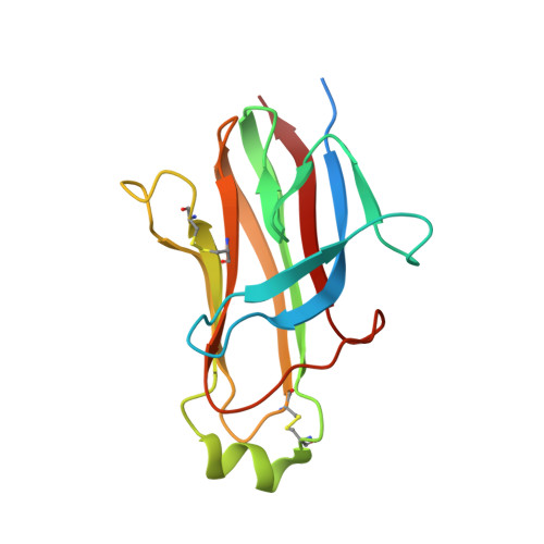
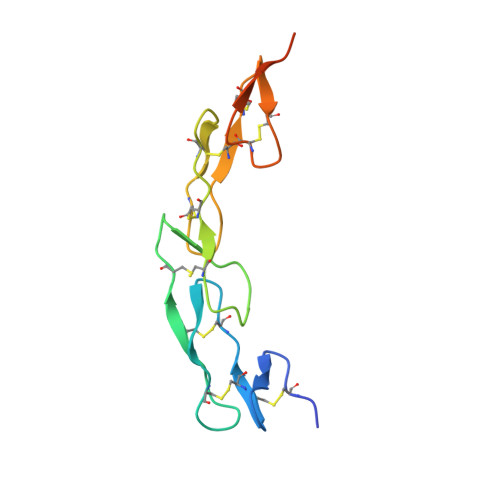
    CD27 is a tumor necrosis factor (TNF) receptor, which stimulates lymphocytes and promotes their differentiation upon activation by TNF ligand CD70. Activation of the CD27 receptor provides a costimulatory signal to promote T cell, B cell, and NK cell activity to facilitate antitumor and anti-infection immunity. Aberrant increased and focused expression of CD70 on many tumor cells renders CD70 an attractive therapeutic target for direct tumor killing. However, despite their use as drug targets to treat cancers, the molecular basis and atomic details of CD27 and CD70 interaction remain elusive. Here we report the crystal structure of human CD27 in complex with human CD70. Analysis of our structure shows that CD70 adopts a classical TNF ligand homotrimeric assembly to engage CD27 receptors in a 3:3 stoichiometry. By combining structural and rational mutagenesis data with reported disease-correlated mutations, we identified the key amino acid residues of CD27 and CD70 that control this interaction. We also report increased potency for plate-bound CD70 constructs compared with solution-phase ligand in a functional activity to stimulate T-cells in vitro. These findings offer new mechanistic insight into this critical costimulatory interaction.


  • Organizational Affiliation

    Pfizer, Inc, La Jolla, California, USA.


Macromolecules
Find similar proteins by:  (by identity cutoff)  |  3D Structure
Entity ID: 1
MoleculeChains Sequence LengthOrganismDetailsImage
CD70 antigen
A, B, C
158Homo sapiensMutation(s): 0 
Gene Names: CD70CD27LCD27LGTNFSF7
UniProt & NIH Common Fund Data Resources
Find proteins for P32970 (Homo sapiens)
Explore P32970 
Go to UniProtKB:  P32970
PHAROS:  P32970
GTEx:  ENSG00000125726 
Entity Groups  
Sequence Clusters30% Identity50% Identity70% Identity90% Identity95% Identity100% Identity
UniProt GroupP32970
Sequence Annotations
Expand
  • Reference Sequence
Find similar proteins by:  (by identity cutoff)  |  3D Structure
Entity ID: 2
MoleculeChains Sequence LengthOrganismDetailsImage
CD27 antigen
D, E, F
114Homo sapiensMutation(s): 0 
Gene Names: CD27TNFRSF7
UniProt & NIH Common Fund Data Resources
Find proteins for P26842 (Homo sapiens)
Explore P26842 
Go to UniProtKB:  P26842
PHAROS:  P26842
GTEx:  ENSG00000139193 
Entity Groups  
Sequence Clusters30% Identity50% Identity70% Identity90% Identity95% Identity100% Identity
UniProt GroupP26842
Sequence Annotations
Expand
  • Reference Sequence
Oligosaccharides

Help

Entity ID: 3
MoleculeChains Length2D Diagram Glycosylation3D Interactions
beta-D-mannopyranose-(1-4)-2-acetamido-2-deoxy-beta-D-glucopyranose-(1-4)-[alpha-L-fucopyranose-(1-6)]2-acetamido-2-deoxy-beta-D-glucopyranose
G, I
4N-Glycosylation
Glycosylation Resources
GlyTouCan:  G32152BH
GlyCosmos:  G32152BH
GlyGen:  G32152BH
Entity ID: 4
MoleculeChains Length2D Diagram Glycosylation3D Interactions
alpha-D-mannopyranose-(1-3)-beta-D-mannopyranose-(1-4)-2-acetamido-2-deoxy-beta-D-glucopyranose-(1-4)-2-acetamido-2-deoxy-beta-D-glucopyranose
H
4N-Glycosylation
Glycosylation Resources
GlyTouCan:  G81315DD
GlyCosmos:  G81315DD
GlyGen:  G81315DD
Entity ID: 5
MoleculeChains Length2D Diagram Glycosylation3D Interactions
alpha-D-mannopyranose-(1-3)-[alpha-D-mannopyranose-(1-6)]beta-D-mannopyranose-(1-4)-2-acetamido-2-deoxy-beta-D-glucopyranose-(1-4)-2-acetamido-2-deoxy-beta-D-glucopyranose
J, L
5N-Glycosylation
Glycosylation Resources
GlyTouCan:  G22768VO
GlyCosmos:  G22768VO
GlyGen:  G22768VO
Entity ID: 6
MoleculeChains Length2D Diagram Glycosylation3D Interactions
alpha-D-mannopyranose-(1-6)-beta-D-mannopyranose-(1-4)-2-acetamido-2-deoxy-beta-D-glucopyranose-(1-4)-[alpha-L-fucopyranose-(1-6)]2-acetamido-2-deoxy-beta-D-glucopyranose
K
5N-Glycosylation
Glycosylation Resources
GlyTouCan:  G00395TQ
GlyCosmos:  G00395TQ
GlyGen:  G00395TQ
Experimental Data & Validation

Experimental Data

  • Method: X-RAY DIFFRACTION
  • Resolution: 2.69 Å
  • R-Value Free: 0.236 
  • R-Value Work: 0.200 
  • R-Value Observed: 0.202 
  • Space Group: I 2 2 2
Unit Cell:
Length ( Å )Angle ( ˚ )
a = 109.53α = 90
b = 113.63β = 90
c = 163.51γ = 90
Software Package:
Software NamePurpose
BUSTERrefinement
XDSdata reduction
XSCALEdata scaling
STARANISOdata reduction
MOLREPphasing
PDB_EXTRACTdata extraction

Structure Validation

View Full Validation Report


Ligand Structure Quality Assessment 


Entry History 

Deposition Data

Revision History  (Full details and data files)

  • Version 1.0: 2021-09-08
    Type: Initial release
  • Version 1.1: 2021-10-13
    Changes: Database references
  • Version 1.2: 2023-10-18
    Changes: Data collection, Refinement description