7KMD

Crystal structure of a HIV-1 clade C isolate Du172.17 HR1.R4.664 Env trimer in complex with human Fabs PGT124 and 35O22


Experimental Data Snapshot

  • Method: X-RAY DIFFRACTION
  • Resolution: 3.39 Å
  • R-Value Free: 0.292 
  • R-Value Work: 0.242 
  • R-Value Observed: 0.244 

wwPDB Validation   3D Report Full Report


Ligand Structure Quality Assessment 


This is version 1.2 of the entry. See complete history


Literature

Neutralizing Antibodies Induced by First-Generation gp41-Stabilized HIV-1 Envelope Trimers and Nanoparticles.

Kumar, S.Lin, X.Ngo, T.Shapero, B.Sou, C.Allen, J.D.Copps, J.Zhang, L.Ozorowski, G.He, L.Crispin, M.Ward, A.B.Wilson, I.A.Zhu, J.

(2021) mBio 12: e0042921-e0042921

  • DOI: https://doi.org/10.1128/mBio.00429-21
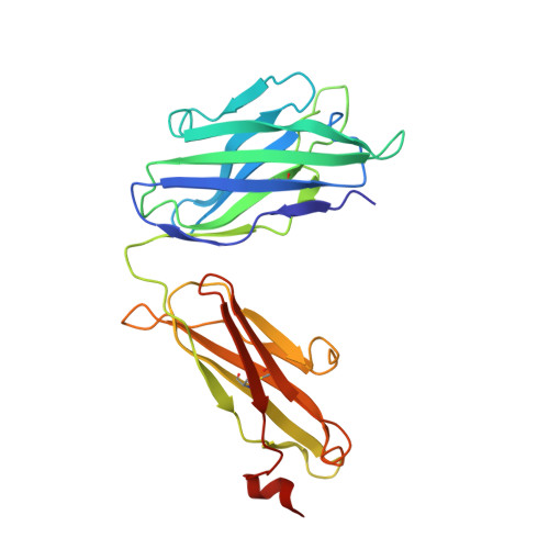
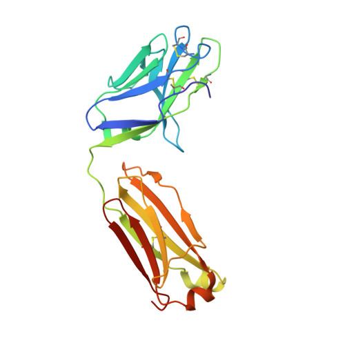
  • Primary Citation of Related Structures:  
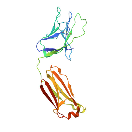
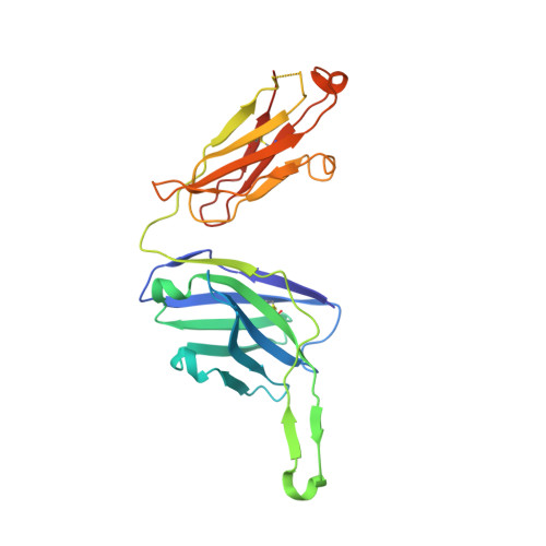
    7KKZ, 7KLC, 7KMD

  • PubMed Abstract: 

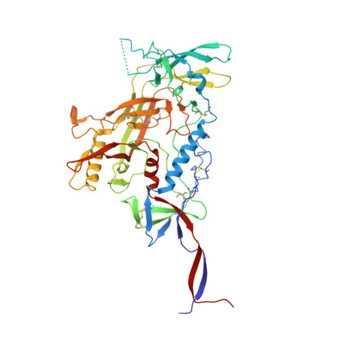
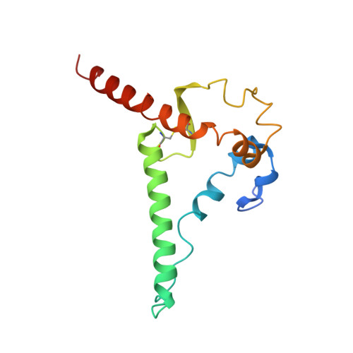
    The immunogenicity of gp41-stabilized HIV-1 BG505 envelope (Env) trimers and nanoparticles (NPs) was recently assessed in mice and rabbits. Here, we combined Env-specific B-cell sorting and repertoire sequencing to identify neutralizing antibodies (NAbs) from immunized animals. A panel of mouse NAbs was isolated from mice immunized with a 60-meric I3-01 NP presenting 20 stabilized trimers. Three mouse NAbs potently neutralized BG505.T332N by recognizing a glycan epitope centered in the C3/V4 region on BG505 Env, as revealed by electron microscopy (EM), X-ray crystallography, and epitope mapping. A set of rabbit NAbs was isolated from rabbits immunized with a soluble trimer and a 24-meric ferritin NP presenting 8 trimers. Neutralization assays against BG505.T332N variants confirmed that potent rabbit NAbs targeted previously described glycan holes on BG505 Env and accounted for a significant portion of the autologous NAb response in both the trimer and ferritin NP groups. Last, we examined NAb responses that were induced by non-BG505 Env immunogens. We determined a 3.4-Å-resolution crystal structure for the clade C transmitted/founder (T/F) Du172.17 Env with a redesigned heptad repeat 1 (HR1) bend in gp41. This clade C Env, in a soluble trimer form and in a multivalent form with 8 trimers attached to ferritin NP, and the gp41-stabilized clade A Q482-d12 Env trimer elicited distinct NAb responses in rabbits, with notable differences in neutralization breadth. Although eliciting a broad NAb response remains a major challenge, our study provides valuable information on an HIV-1 vaccine design strategy that combines gp41 stabilization and NP display. IMPORTANCE Self-assembling protein nanoparticles (NPs) presenting BG505 envelope (Env) trimers can elicit tier 2 HIV-1-neutralizing antibody (NAb) responses more effectively than soluble trimers. In the present study, monoclonal NAbs were isolated from previously immunized mice and rabbits for structural and functional analyses, which revealed that potent mouse NAbs recognize the C3/V4 region and small NP-elicited rabbit NAbs primarily target known glycan holes on BG505 Env. This study validates the gp41 stabilization strategy for HIV-1 Env vaccine design and highlights the challenge in eliciting a broad NAb response.


  • Organizational Affiliation

    Department of Integrative Structural and Computational Biology, The Scripps Research Institutegrid.214007.0, La Jolla, California, USA.


Macromolecules
Find similar proteins by:  (by identity cutoff)  |  3D Structure
Entity ID: 1
MoleculeChains Sequence LengthOrganismDetailsImage
35O22 Heavy chain243Homo sapiensMutation(s): 0 
Entity Groups  
Sequence Clusters30% Identity50% Identity70% Identity90% Identity95% Identity100% Identity
Sequence Annotations
Expand
  • Reference Sequence
Find similar proteins by:  (by identity cutoff)  |  3D Structure
Entity ID: 2
MoleculeChains Sequence LengthOrganismDetailsImage
35O22 Light chainB [auth E]216Homo sapiensMutation(s): 0 
Entity Groups  
Sequence Clusters30% Identity50% Identity70% Identity90% Identity95% Identity100% Identity
Sequence Annotations
Expand
  • Reference Sequence
Find similar proteins by:  (by identity cutoff)  |  3D Structure
Entity ID: 3
MoleculeChains Sequence LengthOrganismDetailsImage
Envelope glycoprotein gp120C [auth G]480Human immunodeficiency virus 1Mutation(s): 0 
Gene Names: env
UniProt
Find proteins for Q202J8 (Human immunodeficiency virus 1)
Explore Q202J8 
Go to UniProtKB:  Q202J8
Entity Groups  
Sequence Clusters30% Identity50% Identity70% Identity90% Identity95% Identity100% Identity
UniProt GroupQ202J8
Sequence Annotations
Expand
  • Reference Sequence
Find similar proteins by:  (by identity cutoff)  |  3D Structure
Entity ID: 4
MoleculeChains Sequence LengthOrganismDetailsImage
Envelope glycoprotein gp41D [auth T]140Human immunodeficiency virus 1Mutation(s): 0 
Gene Names: env
Entity Groups  
Sequence Clusters30% Identity50% Identity70% Identity90% Identity95% Identity100% Identity
Sequence Annotations
Expand
  • Reference Sequence
Find similar proteins by:  (by identity cutoff)  |  3D Structure
Entity ID: 5
MoleculeChains Sequence LengthOrganismDetailsImage
124 Light chainE [auth L]214Homo sapiensMutation(s): 0 
Entity Groups  
Sequence Clusters30% Identity50% Identity70% Identity90% Identity95% Identity100% Identity
Sequence Annotations
Expand
  • Reference Sequence
Find similar proteins by:  (by identity cutoff)  |  3D Structure
Entity ID: 6
MoleculeChains Sequence LengthOrganismDetailsImage
124 Heavy chainF [auth H]236Homo sapiensMutation(s): 0 
Entity Groups  
Sequence Clusters30% Identity50% Identity70% Identity90% Identity95% Identity100% Identity
Sequence Annotations
Expand
  • Reference Sequence
Oligosaccharides

Help

Entity ID: 7
MoleculeChains Length2D Diagram Glycosylation3D Interactions
alpha-D-mannopyranose-(1-2)-alpha-D-mannopyranose-(1-6)-[alpha-D-mannopyranose-(1-3)]alpha-D-mannopyranose-(1-6)-[alpha-D-mannopyranose-(1-2)-alpha-D-mannopyranose-(1-3)]beta-D-mannopyranose-(1-4)-2-acetamido-2-deoxy-beta-D-glucopyranose-(1-4)-2-acetamido-2-deoxy-beta-D-glucopyranoseG [auth B]9N-Glycosylation
Glycosylation Resources
GlyTouCan:  G83161QT
GlyCosmos:  G83161QT
GlyGen:  G83161QT
Entity ID: 8
MoleculeChains Length2D Diagram Glycosylation3D Interactions
2-acetamido-2-deoxy-beta-D-glucopyranose-(1-4)-2-acetamido-2-deoxy-beta-D-glucopyranoseH [auth C],
J [auth F],
O [auth N]
2N-Glycosylation
Glycosylation Resources
GlyTouCan:  G42666HT
GlyCosmos:  G42666HT
GlyGen:  G42666HT
Entity ID: 9
MoleculeChains Length2D Diagram Glycosylation3D Interactions
beta-D-mannopyranose-(1-4)-2-acetamido-2-deoxy-beta-D-glucopyranose-(1-4)-2-acetamido-2-deoxy-beta-D-glucopyranoseI [auth D],
N [auth M],
P [auth O],
Q [auth P]
3N-Glycosylation
Glycosylation Resources
GlyTouCan:  G15407YE
GlyCosmos:  G15407YE
GlyGen:  G15407YE
Entity ID: 10
MoleculeChains Length2D Diagram Glycosylation3D Interactions
alpha-D-mannopyranose-(1-2)-alpha-D-mannopyranose-(1-2)-alpha-D-mannopyranose-(1-3)-[alpha-D-mannopyranose-(1-3)-[alpha-D-mannopyranose-(1-6)]alpha-D-mannopyranose-(1-6)]beta-D-mannopyranose-(1-4)-2-acetamido-2-deoxy-beta-D-glucopyranose-(1-4)-2-acetamido-2-deoxy-beta-D-glucopyranoseK [auth I]9N-Glycosylation
Glycosylation Resources
GlyTouCan:  G68668TB
GlyCosmos:  G68668TB
GlyGen:  G68668TB
Entity ID: 11
MoleculeChains Length2D Diagram Glycosylation3D Interactions
alpha-D-mannopyranose-(1-3)-[alpha-D-mannopyranose-(1-6)]beta-D-mannopyranose-(1-4)-2-acetamido-2-deoxy-beta-D-glucopyranose-(1-4)-2-acetamido-2-deoxy-beta-D-glucopyranoseL [auth J]5N-Glycosylation
Glycosylation Resources
GlyTouCan:  G22768VO
GlyCosmos:  G22768VO
GlyGen:  G22768VO
Entity ID: 12
MoleculeChains Length2D Diagram Glycosylation3D Interactions
alpha-D-mannopyranose-(1-3)-beta-D-mannopyranose-(1-4)-2-acetamido-2-deoxy-beta-D-glucopyranose-(1-4)-2-acetamido-2-deoxy-beta-D-glucopyranose-(6-8)-[2-acetamido-2-deoxy-beta-D-glucopyranose-(1-4)]2-acetamido-2-deoxy-beta-D-glucopyranoseM [auth K]6N/AN-Glycosylation
Glycosylation Resources
GlyTouCan:  G66602QH
GlyCosmos:  G66602QH
Experimental Data & Validation

Experimental Data

  • Method: X-RAY DIFFRACTION
  • Resolution: 3.39 Å
  • R-Value Free: 0.292 
  • R-Value Work: 0.242 
  • R-Value Observed: 0.244 
  • Space Group: P 63
Unit Cell:
Length ( Å )Angle ( ˚ )
a = 127.016α = 90
b = 127.016β = 90
c = 316.538γ = 120
Software Package:
Software NamePurpose
PHENIXrefinement
PHASERphasing
Cootmodel building
HKL-2000data scaling
HKL-2000data reduction

Structure Validation

View Full Validation Report


Ligand Structure Quality Assessment 


Entry History & Funding Information

Deposition Data


Funding OrganizationLocationGrant Number
National Institutes of Health/National Institute Of Allergy and Infectious Diseases (NIH/NIAID)United States--

Revision History  (Full details and data files)

  • Version 1.0: 2021-07-07
    Type: Initial release
  • Version 1.1: 2021-07-28
    Changes: Database references
  • Version 1.2: 2023-10-18
    Changes: Advisory, Data collection, Database references, Refinement description