7K5C

Structure of T7 DNA ejectosome periplasmic tunnel


Experimental Data Snapshot

  • Method: ELECTRON MICROSCOPY
  • Resolution: 2.70 Å
  • Aggregation State: PARTICLE 
  • Reconstruction Method: SINGLE PARTICLE 

wwPDB Validation   3D Report Full Report


This is version 1.1 of the entry. See complete history


Literature

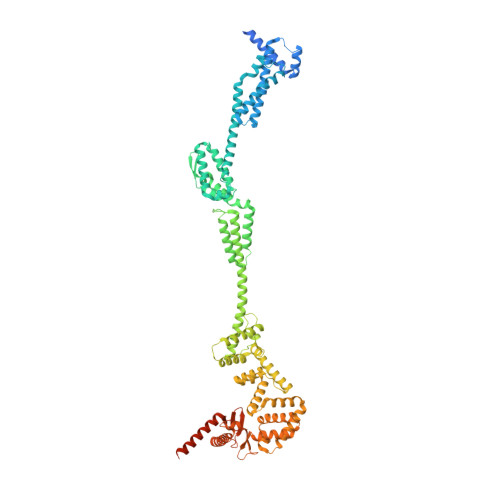
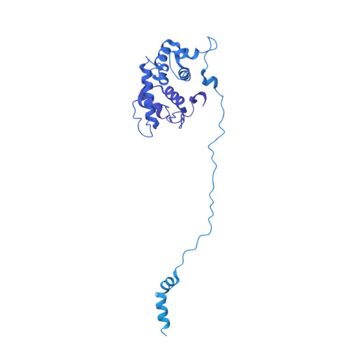
Cryo-EM structure of the periplasmic tunnel of T7 DNA-ejectosome at 2.7 angstrom resolution.

Swanson, N.A.Lokareddy, R.K.Li, F.Hou, C.D.Leptihn, S.Pavlenok, M.Niederweis, M.Pumroy, R.A.Moiseenkova-Bell, V.Y.Cingolani, G.

(2021) Mol Cell 81: 3145

  • DOI: https://doi.org/10.1016/j.molcel.2021.06.001
  • Primary Citation of Related Structures:  
    7K5C

  • PubMed Abstract: 

    Hershey and Chase used bacteriophage T2 genome delivery inside Escherichia coli to demonstrate that DNA, not protein, is the genetic material. Seventy years later, our understanding of viral genome delivery in prokaryotes remains limited, especially for short-tailed phages of the Podoviridae family. These viruses expel mysterious ejection proteins found inside the capsid to form a DNA-ejectosome for genome delivery into bacteria. Here, we reconstitute the phage T7 DNA-ejectosome components gp14, gp15, and gp16 and solve the periplasmic tunnel structure at 2.7 Å resolution. We find that gp14 forms an outer membrane pore, gp15 assembles into a 210 Å hexameric DNA tube spanning the host periplasm, and gp16 extends into the host cytoplasm forming a ∼4,200 residue hub. Gp16 promotes gp15 oligomerization, coordinating peptidoglycan hydrolysis, DNA binding, and lipid insertion. The reconstituted gp15:gp16 complex lacks channel-forming activity, suggesting that the pore for DNA passage forms only transiently during genome ejection.


  • Organizational Affiliation

    Department of Biochemistry and Molecular Biology, Thomas Jefferson University, 1020 Locust Street, Philadelphia, PA 19107, USA.


Macromolecules
Find similar proteins by:  (by identity cutoff)  |  3D Structure
Entity ID: 1
MoleculeChains Sequence LengthOrganismDetailsImage
Internal virion protein gp15747Escherichia phage T7Mutation(s): 0 
Gene Names: 15
UniProt
Find proteins for P03725 (Escherichia phage T7)
Explore P03725 
Go to UniProtKB:  P03725
Entity Groups  
Sequence Clusters30% Identity50% Identity70% Identity90% Identity95% Identity100% Identity
UniProt GroupP03725
Sequence Annotations
Expand
  • Reference Sequence
Find similar proteins by:  (by identity cutoff)  |  3D Structure
Entity ID: 2
MoleculeChains Sequence LengthOrganismDetailsImage
Peptidoglycan transglycosylase gp161,318Escherichia phage T7Mutation(s): 0 
Gene Names: 16
EC: 4.2.2
UniProt
Find proteins for P03726 (Escherichia phage T7)
Explore P03726 
Go to UniProtKB:  P03726
Entity Groups  
Sequence Clusters30% Identity50% Identity70% Identity90% Identity95% Identity100% Identity
UniProt GroupP03726
Sequence Annotations
Expand
  • Reference Sequence
Experimental Data & Validation

Experimental Data

  • Method: ELECTRON MICROSCOPY
  • Resolution: 2.70 Å
  • Aggregation State: PARTICLE 
  • Reconstruction Method: SINGLE PARTICLE 
EM Software:
TaskSoftware PackageVersion
RECONSTRUCTIONRELION3.0-b
MODEL REFINEMENTPHENIX1.17

Structure Validation

View Full Validation Report


Entry History & Funding Information

Deposition Data


Funding OrganizationLocationGrant Number
National Institutes of Health/National Institute of General Medical Sciences (NIH/NIGMS)United StatesGM100888

Revision History  (Full details and data files)

  • Version 1.0: 2021-07-28
    Type: Initial release
  • Version 1.1: 2021-08-18
    Changes: Database references