7JNI

Crystal structure of the angiotensin II type 2 receptoror (AT2R) in complex with EMA401


Experimental Data Snapshot

  • Method: X-RAY DIFFRACTION
  • Resolution: 3.00 Å
  • R-Value Free: 0.273 
  • R-Value Work: 0.234 
  • R-Value Observed: 0.236 

wwPDB Validation   3D Report Full Report


Ligand Structure Quality Assessment 


This is version 1.2 of the entry. See complete history


Literature

Inhibition of the angiotensin II type 2 receptor AT 2 R is a novel therapeutic strategy for glioblastoma.

Perryman, R.Renziehausen, A.Shaye, H.Kostagianni, A.D.Tsiailanis, A.D.Thorne, T.Chatziathanasiadou, M.V.Sivolapenko, G.B.El Mubarak, M.A.Han, G.W.Zarzycka, B.Katritch, V.Lebon, G.Lo Nigro, C.Lattanzio, L.Morse, S.V.Choi, J.J.O'Neill, K.Kanaki, Z.Klinakis, A.Crook, T.Cherezov, V.Tzakos, A.G.Syed, N.

(2022) Proc Natl Acad Sci U S A 119: e2116289119-e2116289119

  • DOI: https://doi.org/10.1073/pnas.2116289119
  • Primary Citation of Related Structures:  
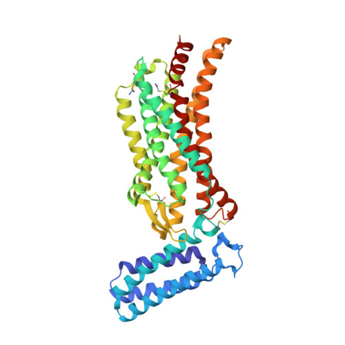
    7JNI

  • PubMed Abstract: 

    Glioblastoma (GBM) is an aggressive malignant primary brain tumor with limited therapeutic options. We show that the angiotensin II (AngII) type 2 receptor (AT 2 R) is a therapeutic target for GBM and that AngII, endogenously produced in GBM cells, promotes proliferation through AT 2 R. We repurposed EMA401, an AT 2 R antagonist originally developed as a peripherally restricted analgesic, for GBM and showed that it inhibits the proliferation of AT 2 R-expressing GBM spheroids and blocks their invasiveness and angiogenic capacity. The crystal structure of AT 2 R bound to EMA401 was determined and revealed the receptor to be in an active-like conformation with helix-VIII blocking G-protein or β-arrestin recruitment. The architecture and interactions of EMA401 in AT 2 R differ drastically from complexes of AT 2 R with other relevant compounds. To enhance central nervous system (CNS) penetration of EMA401, we exploited the crystal structure to design an angiopep-2-tethered EMA401 derivative, A3E. A3E exhibited enhanced CNS penetration, leading to reduced tumor volume, inhibition of proliferation, and increased levels of apoptosis in an orthotopic xenograft model of GBM.


  • Organizational Affiliation

    John Fulcher Molecular Neuro-Oncology Laboratory, Department Brain Sciences, Imperial College, London, United Kingdom.


Macromolecules
Find similar proteins by:  (by identity cutoff)  |  3D Structure
Entity ID: 1
MoleculeChains Sequence LengthOrganismDetailsImage
Soluble cytochrome b562,Type-2 angiotensin II receptor
A, B
412Escherichia coliHomo sapiens
This entity is chimeric
Mutation(s): 3 
Gene Names: cybCAGTR2
Membrane Entity: Yes 
UniProt & NIH Common Fund Data Resources
Find proteins for P0ABE7 (Escherichia coli)
Explore P0ABE7 
Go to UniProtKB:  P0ABE7
Find proteins for P50052 (Homo sapiens)
Explore P50052 
Go to UniProtKB:  P50052
PHAROS:  P50052
GTEx:  ENSG00000180772 
Entity Groups  
Sequence Clusters30% Identity50% Identity70% Identity90% Identity95% Identity100% Identity
UniProt GroupsP0ABE7P50052
Sequence Annotations
Expand
  • Reference Sequence
Small Molecules
Ligands 5 Unique
IDChains Name / Formula / InChI Key2D Diagram3D Interactions
VFD (Subject of Investigation/LOI)
Query on VFD

Download Ideal Coordinates CCD File 
C [auth A],
L [auth B]
Olodanrigan
C32 H29 N O5
GHBCIXGRCZIPNQ-MHZLTWQESA-N
OLC
Query on OLC

Download Ideal Coordinates CCD File 
D [auth A],
G [auth A],
H [auth A]
(2R)-2,3-dihydroxypropyl (9Z)-octadec-9-enoate
C21 H40 O4
RZRNAYUHWVFMIP-GDCKJWNLSA-N
OLA
Query on OLA

Download Ideal Coordinates CCD File 
E [auth A],
F [auth A],
I [auth A],
M [auth B]
OLEIC ACID
C18 H34 O2
ZQPPMHVWECSIRJ-KTKRTIGZSA-N
HEZ
Query on HEZ

Download Ideal Coordinates CCD File 
K [auth A]HEXANE-1,6-DIOL
C6 H14 O2
XXMIOPMDWAUFGU-UHFFFAOYSA-N
FMT
Query on FMT

Download Ideal Coordinates CCD File 
J [auth A]FORMIC ACID
C H2 O2
BDAGIHXWWSANSR-UHFFFAOYSA-N
Modified Residues  1 Unique
IDChains TypeFormula2D DiagramParent
YCM
Query on YCM
A, B
L-PEPTIDE LINKINGC5 H10 N2 O3 SCYS
Experimental Data & Validation

Experimental Data

  • Method: X-RAY DIFFRACTION
  • Resolution: 3.00 Å
  • R-Value Free: 0.273 
  • R-Value Work: 0.234 
  • R-Value Observed: 0.236 
  • Space Group: P 1 21 1
Unit Cell:
Length ( Å )Angle ( ˚ )
a = 77.658α = 90
b = 68.09β = 104.65
c = 89.171γ = 90
Software Package:
Software NamePurpose
PHENIXrefinement
PDB_EXTRACTdata extraction
HKL-2000data reduction
HKL-2000data scaling
PHASERphasing

Structure Validation

View Full Validation Report


Ligand Structure Quality Assessment 


Entry History 

Deposition Data

Revision History  (Full details and data files)

  • Version 1.0: 2022-02-09
    Type: Initial release
  • Version 1.1: 2022-08-24
    Changes: Database references
  • Version 1.2: 2023-10-18
    Changes: Data collection, Refinement description