7EKW

Crystal Structure of the Candida Glabrata Glycogen Debranching Enzyme (D535N) in complex with maltotetrose


Experimental Data Snapshot

  • Method: X-RAY DIFFRACTION
  • Resolution: 3.10 Å
  • R-Value Free: 0.272 
  • R-Value Work: 0.248 
  • R-Value Observed: 0.249 

wwPDB Validation   3D Report Full Report


This is version 1.1 of the entry. See complete history


Literature

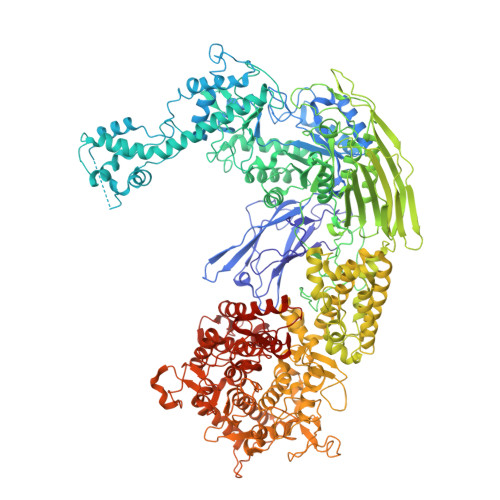
Crystal structures of glycogen-debranching enzyme mutants in complex with oligosaccharides.

Shen, M.Gong, X.Xiang, S.

(2021) Acta Crystallogr F Struct Biol Commun 77: 420-426

  • DOI: https://doi.org/10.1107/S2053230X21010918
  • Primary Citation of Related Structures:  
    7EIM, 7EJP, 7EJT, 7EKW, 7EKX

  • PubMed Abstract: 

    Debranching is a critical step in the mobilization of the important energy store glycogen. In eukaryotes, including fungi and animals, the highly conserved glycogen-debranching enzyme (GDE) debranches glycogen by a glucanotransferase (GT) reaction followed by a glucosidase (GC) reaction. Previous work indicated that these reactions are catalyzed by two active sites located more than 50 Å apart and provided insights into their catalytic mechanisms and substrate recognition. Here, five crystal structures of GDE in complex with oligosaccharides with 4-9 glucose residues are presented. The data suggest that the glycogen main chain plays a critical role in binding to the GT and GC active sites of GDE and that a minimum of five main-chain residues are required for optimal binding.


  • Organizational Affiliation

    Department of Biochemistry and Molecular Biology, Key Laboratory of Immune Microenvironment and Disease (Ministry of Education), The Province and Ministry Co-sponsored Collaborative Innovation Center for Medical Epigenetics, Tianjin Medical University, 22 Qixiangtai Road, Heping District, Tianjin 300070, People's Republic of China.


Macromolecules
Find similar proteins by:  (by identity cutoff)  |  3D Structure
Entity ID: 1
MoleculeChains Sequence LengthOrganismDetailsImage
4-alpha-glucanotransferase
A, B
1,536Nakaseomyces glabratus CBS 138Mutation(s): 1 
Gene Names: GDB1CAGL0G09977g
EC: 2.4.1.25 (PDB Primary Data), 3.2.1.33 (PDB Primary Data)
UniProt
Find proteins for Q6FSK0 (Candida glabrata (strain ATCC 2001 / BCRC 20586 / JCM 3761 / NBRC 0622 / NRRL Y-65 / CBS 138))
Explore Q6FSK0 
Go to UniProtKB:  Q6FSK0
Entity Groups  
Sequence Clusters30% Identity50% Identity70% Identity90% Identity95% Identity100% Identity
UniProt GroupQ6FSK0
Sequence Annotations
Expand
  • Reference Sequence
Oligosaccharides

Help

Entity ID: 2
MoleculeChains Length2D Diagram Glycosylation3D Interactions
alpha-D-glucopyranose-(1-4)-alpha-D-glucopyranose
C
2N/A
Glycosylation Resources
GlyTouCan:  G07411ON
GlyCosmos:  G07411ON
Experimental Data & Validation

Experimental Data

  • Method: X-RAY DIFFRACTION
  • Resolution: 3.10 Å
  • R-Value Free: 0.272 
  • R-Value Work: 0.248 
  • R-Value Observed: 0.249 
  • Space Group: C 1 2 1
Unit Cell:
Length ( Å )Angle ( ˚ )
a = 160.25α = 90
b = 198.94β = 104.87
c = 134.59γ = 90
Software Package:
Software NamePurpose
PHENIXrefinement
PDB_EXTRACTdata extraction
MOSFLMdata reduction
SCALAdata scaling
PHASERphasing

Structure Validation

View Full Validation Report


Entry History & Funding Information

Deposition Data


Funding OrganizationLocationGrant Number
National Natural Science Foundation of China (NSFC)China31870769
National Natural Science Foundation of China (NSFC)China32071205

Revision History  (Full details and data files)

  • Version 1.0: 2021-11-10
    Type: Initial release
  • Version 1.1: 2023-11-29
    Changes: Data collection, Refinement description