7E9S

Archaeal oligosaccharyltransferase AglB from Archaeoglobus fulgidus in complex with an inhibitory peptide and a dolichol-phosphate


Experimental Data Snapshot

  • Method: X-RAY DIFFRACTION
  • Resolution: 2.70 Å
  • R-Value Free: 0.252 
  • R-Value Work: 0.215 
  • R-Value Observed: 0.217 

wwPDB Validation   3D Report Full Report


Ligand Structure Quality Assessment 


This is version 1.1 of the entry. See complete history


Literature

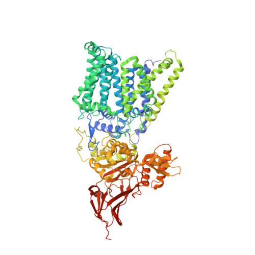
The structure of an archaeal oligosaccharyltransferase provides insight into the strict exclusion of proline from the N-glycosylation sequon.

Taguchi, Y.Yamasaki, T.Ishikawa, M.Kawasaki, Y.Yukimura, R.Mitani, M.Hirata, K.Kohda, D.

(2021) Commun Biol 4: 941-941

  • DOI: https://doi.org/10.1038/s42003-021-02473-8
  • Primary Citation of Related Structures:  
    7E9S

  • PubMed Abstract: 

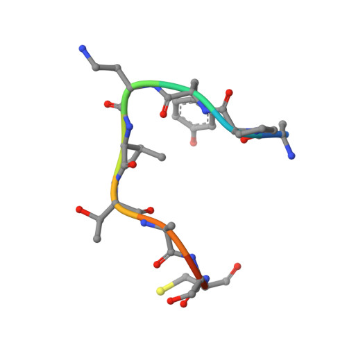
    Oligosaccharyltransferase (OST) catalyzes oligosaccharide transfer to the Asn residue in the N-glycosylation sequon, Asn-X-Ser/Thr, where Pro is strictly excluded at position X. Considering the unique structural properties of proline, this exclusion may not be surprising, but the structural basis for the rejection of Pro residues should be explained explicitly. Here we determined the crystal structure of an archaeal OST in a complex with a sequon-containing peptide and dolichol-phosphate to a 2.7 Å resolution. The sequon part in the peptide forms two inter-chain hydrogen bonds with a conserved amino acid motif, TIXE. We confirmed the essential role of the TIXE motif and the adjacent regions by extensive alanine-scanning of the external loop 5. A Ramachandran plot revealed that the ring structure of the Pro side chain is incompatible with the ϕ backbone dihedral angle around -150° in the rigid sequon-TIXE structure. The present structure clearly provides the structural basis for the exclusion of Pro residues from the N-glycosylation sequon.


  • Organizational Affiliation

    Division of Structural Biology, Medical Institute of Bioregulation, Kyushu University, Fukuoka, Japan.


Macromolecules
Find similar proteins by:  (by identity cutoff)  |  3D Structure
Entity ID: 1
MoleculeChains Sequence LengthOrganismDetailsImage
Dolichyl-phosphooligosaccharide-protein glycotransferase 3875Archaeoglobus fulgidus DSM 4304Mutation(s): 1 
Gene Names: aglB3AF_0380
EC: 2.4.99.21
Membrane Entity: Yes 
UniProt
Find proteins for O29867 (Archaeoglobus fulgidus (strain ATCC 49558 / DSM 4304 / JCM 9628 / NBRC 100126 / VC-16))
Explore O29867 
Go to UniProtKB:  O29867
Entity Groups  
Sequence Clusters30% Identity50% Identity70% Identity90% Identity95% Identity100% Identity
UniProt GroupO29867
Sequence Annotations
Expand
  • Reference Sequence

Find similar proteins by:  Sequence   |   3D Structure  

Entity ID: 2
MoleculeChains Sequence LengthOrganismDetailsImage
a polypeptide linked to an inhibitory N-glycosylation sequon-containing peptide10synthetic constructMutation(s): 0 
Entity Groups  
Sequence Clusters30% Identity50% Identity70% Identity90% Identity95% Identity100% Identity
Sequence Annotations
Expand
  • Reference Sequence
Small Molecules
Ligands 4 Unique
IDChains Name / Formula / InChI Key2D Diagram3D Interactions
J06 (Subject of Investigation/LOI)
Query on J06

Download Ideal Coordinates CCD File 
D [auth A][(3S,6Z,10Z,14Z,18Z,22Z,26Z,30Z,34Z,39S,43S)-3,7,11,15,19,23,27,31,35,39,43,47-dodecamethyloctatetraconta-6,10,14,18,22,26,30,34-octaenyl] dihydrogen phosphate
C60 H107 O4 P
XTTHNPQIJYVHIB-WHVVPYAWSA-N
7E8
Query on 7E8

Download Ideal Coordinates CCD File 
J [auth A],
K [auth A],
L [auth A],
M [auth A]
(2R)-2,3-DIHYDROXYPROPYL (7Z)-TETRADEC-7-ENOATE
C17 H32 O4
LVBAGTJIDOCNIJ-XITLMJRVSA-N
PEG
Query on PEG

Download Ideal Coordinates CCD File 
E [auth A],
F [auth A],
G [auth A],
H [auth A],
I [auth A]
DI(HYDROXYETHYL)ETHER
C4 H10 O3
MTHSVFCYNBDYFN-UHFFFAOYSA-N
MN
Query on MN

Download Ideal Coordinates CCD File 
C [auth A]MANGANESE (II) ION
Mn
WAEMQWOKJMHJLA-UHFFFAOYSA-N
Modified Residues  1 Unique
IDChains TypeFormula2D DiagramParent
DAB
Query on DAB
B
L-PEPTIDE LINKINGC4 H10 N2 O2ALA
Experimental Data & Validation

Experimental Data

  • Method: X-RAY DIFFRACTION
  • Resolution: 2.70 Å
  • R-Value Free: 0.252 
  • R-Value Work: 0.215 
  • R-Value Observed: 0.217 
  • Space Group: P 21 21 2
Unit Cell:
Length ( Å )Angle ( ˚ )
a = 345.74α = 90
b = 48.69β = 90
c = 63.56γ = 90
Software Package:
Software NamePurpose
XDSdata reduction
XSCALEdata scaling
PHENIXphasing
PHENIXrefinement
Cootmodel building

Structure Validation

View Full Validation Report


Ligand Structure Quality Assessment 


Entry History & Funding Information

Deposition Data


Funding OrganizationLocationGrant Number
Japan Agency for Medical Research and Development (AMED)JapanJP19am0101070
Japan Society for the Promotion of Science (JSPS)JapanJP20H00405

Revision History  (Full details and data files)

  • Version 1.0: 2021-09-08
    Type: Initial release
  • Version 1.1: 2023-11-29
    Changes: Data collection, Refinement description