7DED

Mevo lectin complex with mannoheptose (Man7)


Experimental Data Snapshot

  • Method: X-RAY DIFFRACTION
  • Resolution: 2.23 Å
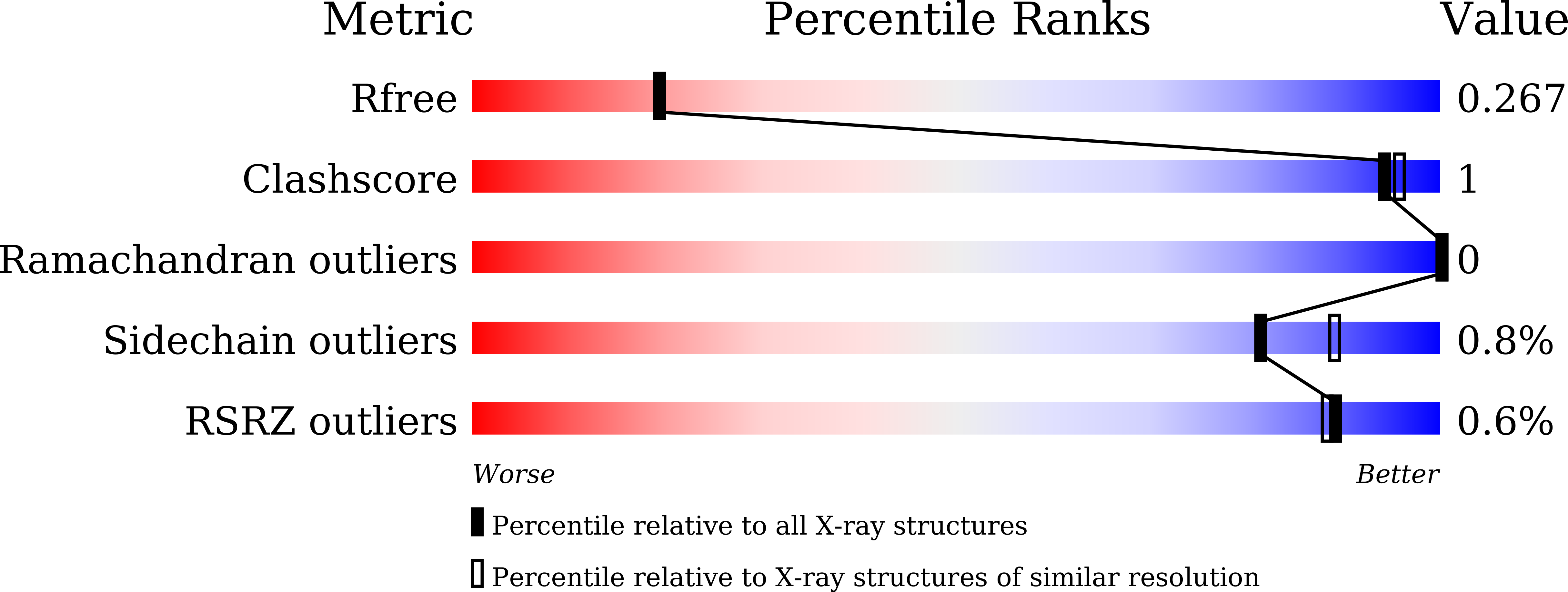
  • R-Value Free: 0.267 
  • R-Value Work: 0.246 

wwPDB Validation   3D Report Full Report


Ligand Structure Quality Assessment 


This is version 1.2 of the entry. See complete history


Literature

Mevo lectin specificity toward high-mannose structures with terminal alpha Man(1,2) alpha Man residues and its implication to inhibition of the entry of Mycobacterium tuberculosis into macrophages.

Sivaji, N.Harish, N.Singh, S.Singh, A.Vijayan, M.Surolia, A.

(2021) Glycobiology 31: 1046-1059

  • DOI: https://doi.org/10.1093/glycob/cwab022
  • Primary Citation of Related Structures:  
    7DED

  • PubMed Abstract: 

    Mannose-binding lectins can specifically recognize and bind complex glycan structures on pathogens and have potential as antiviral and antibacterial agents. We previously reported the structure of a lectin from an archaeal species, Mevo lectin, which has specificity toward terminal α1,2 linked manno-oligosaccharides. Mycobacterium tuberculosis expresses mannosylated structures including lipoarabinomannan (ManLAM) on its surface and exploits C-type lectins to gain entry into the host cells. ManLAM structure has mannose capping with terminal αMan(1,2)αMan residues and is important for recognition by innate immune cells. Here, we aim to address the specificity of Mevo lectin toward high-mannose type glycans with terminal αMan(1,2)αMan residues and its effect on M. tuberculosis internalization by macrophages. Isothermal titration calorimetry studies demonstrated that Mevo lectin shows preferential binding toward manno-oligosaccharides with terminal αMan(1,2)αMan structures and showed a strong affinity for ManLAM, whereas it binds weakly to Mycobacterium smegmatis lipoarabinomannan, which displays relatively fewer and shorter mannosyl caps. Crystal structure of Mevo lectin complexed with a Man7D1 revealed the multivalent cross-linking interaction, which explains avidity-based high-affinity for these ligands when compared to previously studied manno-oligosaccharides lacking the specific termini. Functional studies suggest that M. tuberculosis internalization by the macrophage was impaired by binding of Mevo lectin to ManLAM present on the surface of M. tuberculosis. Selectivity shown by Mevo lectin toward glycans with terminal αMan(1,2)αMan structures, and its ability to compromise the internalization of M. tuberculosis  in vitro, underscore the potential utility of Mevo lectin as a research tool to study host-pathogen interactions.


  • Organizational Affiliation

    Molecular Biophysics Unit, Indian Institute of Science, Bangalore 560012, India.


Macromolecules
Find similar proteins by:  (by identity cutoff)  |  3D Structure
Entity ID: 1
MoleculeChains Sequence LengthOrganismDetailsImage
lectin140Methanococcus voltae A3Mutation(s): 0 
Gene Names: Mvol_0737
UniProt
Find proteins for D7DTD6 (Methanococcus voltae (strain ATCC BAA-1334 / A3))
Explore D7DTD6 
Go to UniProtKB:  D7DTD6
Entity Groups  
Sequence Clusters30% Identity50% Identity70% Identity90% Identity95% Identity100% Identity
UniProt GroupD7DTD6
Sequence Annotations
Expand
  • Reference Sequence
Oligosaccharides

Help

Entity ID: 2
MoleculeChains Length2D Diagram Glycosylation3D Interactions
alpha-D-mannopyranose-(1-2)-alpha-D-mannopyranose-(1-2)-alpha-D-mannopyranose-(1-3)-[alpha-D-mannopyranose-(1-3)-alpha-D-mannopyranose-(1-6)]alpha-D-mannopyranose
H, I, J
6N/A
Glycosylation Resources
GlyTouCan:  G05929XL
GlyCosmos:  G05929XL
GlyGen:  G05929XL
Small Molecules
Ligands 2 Unique
IDChains Name / Formula / InChI Key2D Diagram3D Interactions
MAN (Subject of Investigation/LOI)
Query on MAN

Download Ideal Coordinates CCD File 
K [auth E]alpha-D-mannopyranose
C6 H12 O6
WQZGKKKJIJFFOK-PQMKYFCFSA-N
EDO
Query on EDO

Download Ideal Coordinates CCD File 
L [auth A]
M [auth B]
N [auth B]
O [auth B]
P [auth B]
L [auth A],
M [auth B],
N [auth B],
O [auth B],
P [auth B],
Q [auth B],
R [auth B],
S [auth B],
T [auth C],
U [auth C],
V [auth G],
W [auth G],
X [auth F],
Y [auth F],
Z [auth F]
1,2-ETHANEDIOL
C2 H6 O2
LYCAIKOWRPUZTN-UHFFFAOYSA-N
Experimental Data & Validation

Experimental Data

  • Method: X-RAY DIFFRACTION
  • Resolution: 2.23 Å
  • R-Value Free: 0.267 
  • R-Value Work: 0.246 
  • Space Group: P 42 21 2
Unit Cell:
Length ( Å )Angle ( ˚ )
a = 168.35α = 90
b = 168.35β = 90
c = 104.96γ = 90
Software Package:
Software NamePurpose
REFMACrefinement
iMOSFLMdata reduction
SCALAdata scaling
PHASERphasing

Structure Validation

View Full Validation Report


Ligand Structure Quality Assessment 


Entry History & Funding Information

Deposition Data


Funding OrganizationLocationGrant Number
Department of Science & Technology (DST, India)IndiaEMR/2016/005205

Revision History  (Full details and data files)

  • Version 1.0: 2021-04-21
    Type: Initial release
  • Version 1.1: 2021-10-06
    Changes: Database references
  • Version 1.2: 2023-11-29
    Changes: Data collection, Derived calculations, Refinement description