7D9C

Alpha-glucosidase from Weissella cibaria BBK-1 bound with maltose


Experimental Data Snapshot

  • Method: X-RAY DIFFRACTION
  • Resolution: 1.36 Å
  • R-Value Free: 0.153 
  • R-Value Work: 0.140 
  • R-Value Observed: 0.140 

wwPDB Validation   3D Report Full Report


Ligand Structure Quality Assessment 


This is version 1.2 of the entry. See complete history


Literature

A GH13 alpha-glucosidase from Weissella cibaria uncommonly acts on short-chain maltooligosaccharides.

Wangpaiboon, K.Laohawuttichai, P.Kim, S.Y.Mori, T.Nakapong, S.Pichyangkura, R.Pongsawasdi, P.Hakoshima, T.Krusong, K.

(2021) Acta Crystallogr D Struct Biol 77: 1064-1076

  • DOI: https://doi.org/10.1107/S205979832100677X
  • Primary Citation of Related Structures:  
    7D9B, 7D9C, 7DCG, 7DCH, 7EHH, 7EHI

  • PubMed Abstract: 

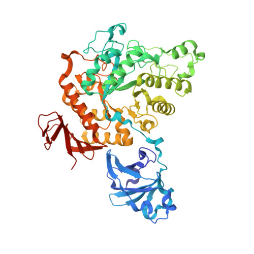
    α-Glucosidase (EC 3.2.1.20) is a carbohydrate-hydrolyzing enzyme which generally cleaves α-1,4-glycosidic bonds of oligosaccharides and starch from the nonreducing ends. In this study, the novel α-glucosidase from Weissella cibaria BBK-1 (WcAG) was biochemically and structurally characterized. WcAG belongs to glycoside hydrolase family 13 (GH13) and to the neopullanase subfamily. It exhibits distinct hydrolytic activity towards the α-1,4 linkages of short-chain oligosaccharides from the reducing end. The enzyme prefers to hydrolyse maltotriose and acarbose, while it cannot hydrolyse cyclic oligosaccharides and polysaccharides. In addition, WcAG can cleave pullulan hydrolysates and strongly exhibits transglycosylation activity in the presence of maltose. Size-exclusion chromatography and X-ray crystal structures revealed that WcAG forms a homodimer in which the N-terminal domain of one monomer is orientated in proximity to the catalytic domain of another, creating the substrate-binding groove. Crystal structures of WcAG in complexes with maltose, maltotriose and acarbose revealed a remarkable enzyme active site with accessible +2, +1 and -1 subsites, along with an Arg-Glu gate (Arg176-Glu296) in front of the active site. The -2 and -3 subsites were blocked by Met119 and Asn120 from the N-terminal domain of a different subunit, resulting in an extremely restricted substrate preference.


  • Organizational Affiliation

    Department of Biochemistry, Faculty of Science, Chulalongkorn University, Bangkok 10330, Thailand.


Macromolecules
Find similar proteins by:  (by identity cutoff)  |  3D Structure
Entity ID: 1
MoleculeChains Sequence LengthOrganismDetailsImage
Alpha-glycosidase589Weissella confusaMutation(s): 0 
Gene Names: C6P08_07940
EC: 3.2.1.20
UniProt
Find proteins for A0A482PRE5 (Weissella confusa)
Explore A0A482PRE5 
Go to UniProtKB:  A0A482PRE5
Entity Groups  
Sequence Clusters30% Identity50% Identity70% Identity90% Identity95% Identity100% Identity
UniProt GroupA0A482PRE5
Sequence Annotations
Expand
  • Reference Sequence
Oligosaccharides

Help

Entity ID: 2
MoleculeChains Length2D Diagram Glycosylation3D Interactions
alpha-D-glucopyranose-(1-4)-alpha-D-glucopyranose
B
2N/A
Glycosylation Resources
GlyTouCan:  G07411ON
GlyCosmos:  G07411ON
Small Molecules
Ligands 5 Unique
IDChains Name / Formula / InChI Key2D Diagram3D Interactions
MES
Query on MES

Download Ideal Coordinates CCD File 
J [auth A],
K [auth A]
2-(N-MORPHOLINO)-ETHANESULFONIC ACID
C6 H13 N O4 S
SXGZJKUKBWWHRA-UHFFFAOYSA-N
BGC (Subject of Investigation/LOI)
Query on BGC

Download Ideal Coordinates CCD File 
M [auth A]beta-D-glucopyranose
C6 H12 O6
WQZGKKKJIJFFOK-VFUOTHLCSA-N
GLC
Query on GLC

Download Ideal Coordinates CCD File 
L [auth A]alpha-D-glucopyranose
C6 H12 O6
WQZGKKKJIJFFOK-DVKNGEFBSA-N
SO4
Query on SO4

Download Ideal Coordinates CCD File 
D [auth A]
E [auth A]
F [auth A]
G [auth A]
H [auth A]
D [auth A],
E [auth A],
F [auth A],
G [auth A],
H [auth A],
I [auth A]
SULFATE ION
O4 S
QAOWNCQODCNURD-UHFFFAOYSA-L
CA (Subject of Investigation/LOI)
Query on CA

Download Ideal Coordinates CCD File 
C [auth A]CALCIUM ION
Ca
BHPQYMZQTOCNFJ-UHFFFAOYSA-N
Biologically Interesting Molecules (External Reference) 1 Unique
Experimental Data & Validation

Experimental Data

  • Method: X-RAY DIFFRACTION
  • Resolution: 1.36 Å
  • R-Value Free: 0.153 
  • R-Value Work: 0.140 
  • R-Value Observed: 0.140 
  • Space Group: P 41 2 2
Unit Cell:
Length ( Å )Angle ( ˚ )
a = 115.636α = 90
b = 115.636β = 90
c = 106.163γ = 90
Software Package:
Software NamePurpose
REFMACrefinement
PHENIXrefinement
DENZOdata reduction
SCALEPACKdata scaling
PHASERphasing

Structure Validation

View Full Validation Report


Ligand Structure Quality Assessment 


Entry History & Funding Information

Deposition Data


Funding OrganizationLocationGrant Number
Other governmentThailand--

Revision History  (Full details and data files)

  • Version 1.0: 2021-08-11
    Type: Initial release
  • Version 1.1: 2021-08-18
    Changes: Database references
  • Version 1.2: 2023-11-29
    Changes: Data collection, Refinement description