7D6A

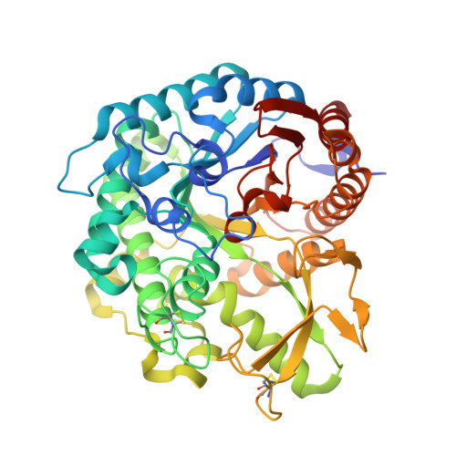
Crystal structure of Oryza sativa Os4BGlu18 monolignol beta-glucosidase


Experimental Data Snapshot

  • Method: X-RAY DIFFRACTION
  • Resolution: 1.70 Å
  • R-Value Free: 0.175 
  • R-Value Work: 0.150 
  • R-Value Observed: 0.151 

Starting Model: experimental
View more details

wwPDB Validation   3D Report Full Report


Ligand Structure Quality Assessment 


This is version 1.2 of the entry. See complete history


Literature

Structural analysis of rice Os4BGlu18 monolignol beta-glucosidase.

Baiya, S.Pengthaisong, S.Kitjaruwankul, S.Ketudat Cairns, J.R.

(2021) PLoS One 16: e0241325-e0241325

  • DOI: https://doi.org/10.1371/journal.pone.0241325
  • Primary Citation of Related Structures:  
    7D6A, 7D6B

  • PubMed Abstract: 

    Monolignol glucosides are storage forms of monolignols, which are polymerized to lignin to strengthen plant cell walls. The conversion of monolignol glucosides to monolignols is catalyzed by monolignol β-glucosidases. Rice Os4BGlu18 β-glucosidase catalyzes hydrolysis of the monolignol glucosides, coniferin, syringin, and p-coumaryl alcohol glucoside more efficiently than other natural substrates. To understand more clearly the basis for substrate specificity of a monolignol β-glucosidase, the structure of Os4BGlu18 was determined by X-ray crystallography. Crystals of Os4BGlu18 and its complex with δ-gluconolactone diffracted to 1.7 and 2.1 Å resolution, respectively. Two protein molecules were found in the asymmetric unit of the P212121 space group of their isomorphous crystals. The Os4BGlu18 structure exhibited the typical (β/α)8 TIM barrel of glycoside hydrolase family 1 (GH1), but the four variable loops and two disulfide bonds appeared significantly different from other known structures of GH1 β-glucosidases. Molecular docking studies of the Os4BGlu18 structure with monolignol substrate ligands placed the glycone in a similar position to the δ-gluconolactone in the complex structure and revealed the interactions between protein and ligands. Molecular docking, multiple sequence alignment, and homology modeling identified amino acid residues at the aglycone-binding site involved in substrate specificity for monolignol β-glucosides. Thus, the structural basis of substrate recognition and hydrolysis by monolignol β-glucosidases was elucidated.


  • Organizational Affiliation

    Faculty of Science at Sriracha, Kasetsart University, Sriracha Campus, Sriracha, Chonburi, Thailand.


Macromolecules
Find similar proteins by:  (by identity cutoff)  |  3D Structure
Entity ID: 1
MoleculeChains Sequence LengthOrganismDetailsImage
Beta-glucosidase 18
A, B
482Oryza sativa Japonica GroupMutation(s): 0 
Gene Names: BGLU18Os04g0513900LOC_Os04g43410OSJNBa0004N05.26OSJNBb0070J16.3
EC: 3.2.1.21
UniProt
Find proteins for Q7XSK0 (Oryza sativa subsp. japonica)
Explore Q7XSK0 
Go to UniProtKB:  Q7XSK0
Entity Groups  
Sequence Clusters30% Identity50% Identity70% Identity90% Identity95% Identity100% Identity
UniProt GroupQ7XSK0
Sequence Annotations
Expand
  • Reference Sequence
Small Molecules
Ligands 3 Unique
IDChains Name / Formula / InChI Key2D Diagram3D Interactions
MES (Subject of Investigation/LOI)
Query on MES

Download Ideal Coordinates CCD File 
C [auth A],
M [auth B]
2-(N-MORPHOLINO)-ETHANESULFONIC ACID
C6 H13 N O4 S
SXGZJKUKBWWHRA-UHFFFAOYSA-N
GOL (Subject of Investigation/LOI)
Query on GOL

Download Ideal Coordinates CCD File 
D [auth A]
E [auth A]
F [auth A]
G [auth A]
H [auth A]
D [auth A],
E [auth A],
F [auth A],
G [auth A],
H [auth A],
I [auth A],
J [auth A],
K [auth A],
N [auth B],
O [auth B],
P [auth B],
Q [auth B],
R [auth B]
GLYCEROL
C3 H8 O3
PEDCQBHIVMGVHV-UHFFFAOYSA-N
ZN (Subject of Investigation/LOI)
Query on ZN

Download Ideal Coordinates CCD File 
L [auth B]ZINC ION
Zn
PTFCDOFLOPIGGS-UHFFFAOYSA-N
Experimental Data & Validation

Experimental Data

  • Method: X-RAY DIFFRACTION
  • Resolution: 1.70 Å
  • R-Value Free: 0.175 
  • R-Value Work: 0.150 
  • R-Value Observed: 0.151 
  • Space Group: P 21 21 21
Unit Cell:
Length ( Å )Angle ( ˚ )
a = 52.134α = 90
b = 83.759β = 90
c = 207.649γ = 90
Software Package:
Software NamePurpose
REFMACrefinement
HKL-3000data reduction
HKL-3000data scaling
MOLREPphasing

Structure Validation

View Full Validation Report


Ligand Structure Quality Assessment 


Entry History & Funding Information

Deposition Data


Funding OrganizationLocationGrant Number
Other governmentThailandBRG5980015 Thailand Research Fund and Suranaree University of Technology
Other governmentThailandSuranaree University of Technology

Revision History  (Full details and data files)

  • Version 1.0: 2021-01-13
    Type: Initial release
  • Version 1.1: 2021-02-10
    Changes: Database references
  • Version 1.2: 2023-11-29
    Changes: Data collection, Database references, Refinement description