Structural Insight into the Contributions of the N-Terminus and Key Active-Site Residues to the Catalytic Efficiency of Glutamine Synthetase 2.
Chen, W.T., Yang, H.Y., Lin, C.Y., Lee, Y.Z., Ma, S.C., Chen, W.C., Yin, H.S.(2020) Biomolecules 10
- PubMed: 33327463
- DOI: https://doi.org/10.3390/biom10121671
- Primary Citation of Related Structures:
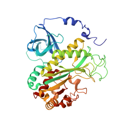
7CPR - PubMed Abstract:
Glutamine synthetase (GS) catalyzes the condensation of ammonia and glutamate, along with ATP, to form glutamine. Despite extensive studies on GSs from eukaryotes and prokaryotes, the roles of the N-terminus and other structural features in catalysis remain unclear. Here we report the decameric structure of Drosophila melanogaster GS 2 (DmGS2). The N-terminal short helices, α1 and α2, constitute a meander region, and form hydrogen bonds with residues 3-5 in the N-terminal loop, which are not present in the GSs of other species. Deletion of α1 or α1-α2 inactivates DmGS2. Notably, the Arg4 in each monomer of one pentamer forms hydrogen bonds with Glu7, and Asp8 in the adjacent monomer of the other pentamer. Replacement of Arg4 with Asp (R4D) abolishes activity. Analytical ultracentrifugation revealed that Arg4 is crucial for oligomerization. Circular dichroism spectra revealed that R4D may alter the secondary structure. We mutated key residues to identify the substrate-binding site. As Glu140 binds glutamate and Glu311 binds ammonia, mutants E140A and E311A have little activity. Conversely, mutant P214A (P contributes to ATP binding) has higher activity than wild-type DmGS2. These findings expand the understanding of the structural and functional features of the N-terminal meander region of DmGS2 and the residues important for catalytic efficiency.
Organizational Affiliation:
Institute of Bioinformatics and Structural Biology, and College of Life Sciences, National Tsing Hua University, No. 101, Section 2, Kuang-Fu Road, Hsinchu 30013, Taiwan.