


Near-perfect control of the regioselective glucosylation enabled by rational design of glycosyltransferases
Li, J., Qu, G., Shang, N., Chen, P., Men, Y., Liu, W.D., Mei, Z., Sun, Y.X., Sun, Z.(2021) Green Synth Catal
Experimental Data Snapshot
Entity ID: 1 | |||||
|---|---|---|---|---|---|
| Molecule | Chains | Sequence Length | Organism | Details | Image |
| Glycosyltransferase | 483 | Siraitia grosvenorii | Mutation(s): 0 Gene Names: UGT74AC2 EC: 2.4.1 | 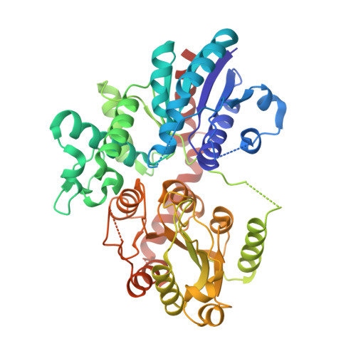 | |
UniProt | |||||
Find proteins for A0A346A6C4 (Siraitia grosvenorii) Explore A0A346A6C4 Go to UniProtKB: A0A346A6C4 | |||||
Entity Groups | |||||
| Sequence Clusters | 30% Identity50% Identity70% Identity90% Identity95% Identity100% Identity | ||||
| UniProt Group | A0A346A6C4 | ||||
Sequence AnnotationsExpand | |||||
| |||||
| Ligands 1 Unique | |||||
|---|---|---|---|---|---|
| ID | Chains | Name / Formula / InChI Key | 2D Diagram | 3D Interactions | |
| UDP (Subject of Investigation/LOI) Query on UDP | C [auth A], D [auth B] | URIDINE-5'-DIPHOSPHATE C9 H14 N2 O12 P2 XCCTYIAWTASOJW-XVFCMESISA-N |  | ||
| Length ( Å ) | Angle ( ˚ ) |
|---|---|
| a = 197.52 | α = 90 |
| b = 63.71 | β = 101.65 |
| c = 93.8 | γ = 90 |
| Software Name | Purpose |
|---|---|
| PHENIX | refinement |
| HKL-2000 | data scaling |
| PDB_EXTRACT | data extraction |
| HKL-2000 | data reduction |
| PHASER | phasing |
RCSB PDB (citation) is hosted by
RCSB PDB is a member of the

