7BOP

Crystal Structure of Core-mannan synthase A (CmsA/Ktr4) from Aspergillus fumigatus, Mn/GDP-form


Experimental Data Snapshot

  • Method: X-RAY DIFFRACTION
  • Resolution: 1.90 Å
  • R-Value Free: 0.181 
  • R-Value Work: 0.160 
  • R-Value Observed: 0.161 

wwPDB Validation   3D Report Full Report


Ligand Structure Quality Assessment 


This is version 1.3 of the entry. See complete history


Literature

Structural basis for the core-mannan biosynthesis of cell wall fungal-type galactomannan in Aspergillus fumigatus .

Hira, D.Onoue, T.Oka, T.

(2020) J Biol Chem 295: 15407-15417

  • DOI: https://doi.org/10.1074/jbc.RA120.013742
  • Primary Citation of Related Structures:  
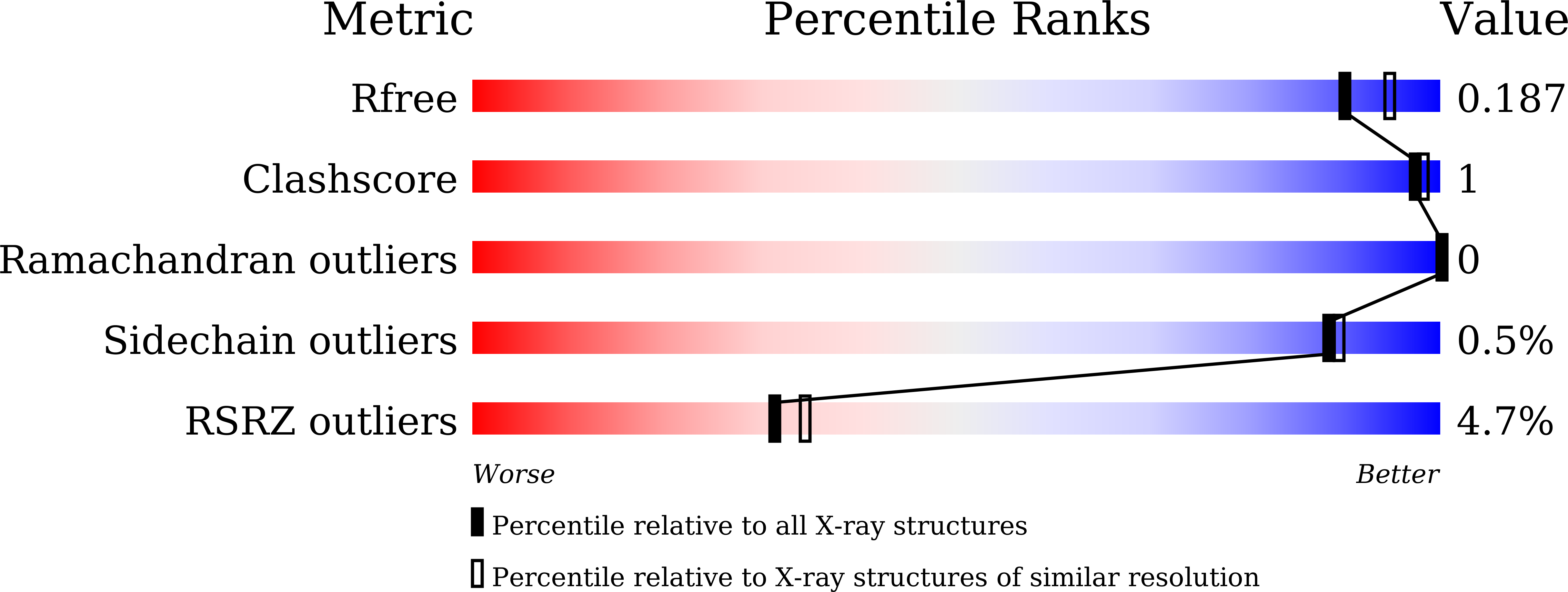
    7BOO, 7BOP

  • PubMed Abstract: 

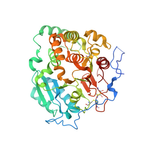
    Fungal cell walls and their biosynthetic enzymes are potential targets for novel antifungal agents. Recently, two mannosyltransferases, namely core-mannan synthases A (CmsA/Ktr4) and B (CmsB/Ktr7), were found to play roles in the core-mannan biosynthesis of fungal-type galactomannan. CmsA/Ktr4 is an α-(1→2)-mannosyltransferase responsible for α-(1→2)-mannan biosynthesis in fungal-type galactomannan, which covers the cell surface of Aspergillus fumigatus Strains with disrupted cmsA / ktr4 have been shown to exhibit strongly suppressed hyphal elongation and conidiation alongside reduced virulence in a mouse model of invasive aspergillosis, indicating that CmsA/Ktr4 is a potential novel antifungal candidate. In this study we present the 3D structures of the soluble catalytic domain of CmsA/Ktr4, as determined by X-ray crystallography at a resolution of 1.95 Å, as well as the enzyme and Mn 2+ /GDP complex to 1.90 Å resolution. The CmsA/Ktr4 protein not only contains a highly conserved binding pocket for the donor substrate, GDP-mannose, but also has a unique broad cleft structure formed by its N- and C-terminal regions and is expected to recognize the acceptor substrate, a mannan chain. Based on these crystal structures, we also present a 3D structural model of the enzyme-substrate complex generated using docking and molecular dynamics simulations with α-Man-(1→6)-α-Man-(1→2)-α-Man-OMe as the model structure for the acceptor substrate. This predicted enzyme-substrate complex structure is also supported by findings from single amino acid substitution CmsA/Ktr4 mutants expressed in Δ cmsA/ktr4 A. fumigatus cells. Taken together, these results provide basic information for developing specific α-mannan biosynthesis inhibitors for use as pharmaceuticals and/or pesticides.


  • Organizational Affiliation

    Department of Applied Life Science, Faculty of Biotechnology and Life Science, Sojo University, Kumamoto, Kumamoto, Japan. Electronic address: hira@life.sojo-u.ac.jp.


Macromolecules
Find similar proteins by:  (by identity cutoff)  |  3D Structure
Entity ID: 1
MoleculeChains Sequence LengthOrganismDetailsImage
Alpha-1,2-mannosyltransferase (Ktr4), putative
A, B, C, D, E
A, B, C, D, E, F
386Aspergillus fumigatus A1163Mutation(s): 0 
Gene Names: AFUB_051270
UniProt
Find proteins for B0Y2F5 (Aspergillus fumigatus (strain CBS 144.89 / FGSC A1163 / CEA10))
Explore B0Y2F5 
Go to UniProtKB:  B0Y2F5
Entity Groups  
Sequence Clusters30% Identity50% Identity70% Identity90% Identity95% Identity100% Identity
UniProt GroupB0Y2F5
Sequence Annotations
Expand
  • Reference Sequence
Small Molecules
Ligands 4 Unique
IDChains Name / Formula / InChI Key2D Diagram3D Interactions
GDP
Query on GDP

Download Ideal Coordinates CCD File 
CA [auth D]
H [auth A]
JA [auth E]
P [auth B]
PA [auth F]
CA [auth D],
H [auth A],
JA [auth E],
P [auth B],
PA [auth F],
X [auth C]
GUANOSINE-5'-DIPHOSPHATE
C10 H15 N5 O11 P2
QGWNDRXFNXRZMB-UUOKFMHZSA-N
GOL (Subject of Investigation/LOI)
Query on GOL

Download Ideal Coordinates CCD File 
AA [auth C]
FA [auth D]
GA [auth D]
HA [auth D]
K [auth A]
AA [auth C],
FA [auth D],
GA [auth D],
HA [auth D],
K [auth A],
KA [auth E],
L [auth A],
LA [auth E],
M [auth A],
MA [auth E],
N [auth A],
NA [auth E],
QA [auth F],
RA [auth F],
S [auth B],
T [auth B],
U [auth B],
V [auth B],
Z [auth C]
GLYCEROL
C3 H8 O3
PEDCQBHIVMGVHV-UHFFFAOYSA-N
MN
Query on MN

Download Ideal Coordinates CCD File 
BA [auth D]
G [auth A]
IA [auth E]
O [auth B]
OA [auth F]
BA [auth D],
G [auth A],
IA [auth E],
O [auth B],
OA [auth F],
W [auth C]
MANGANESE (II) ION
Mn
WAEMQWOKJMHJLA-UHFFFAOYSA-N
NA (Subject of Investigation/LOI)
Query on NA

Download Ideal Coordinates CCD File 
DA [auth D]
EA [auth D]
I [auth A]
J [auth A]
Q [auth B]
DA [auth D],
EA [auth D],
I [auth A],
J [auth A],
Q [auth B],
R [auth B],
Y [auth C]
SODIUM ION
Na
FKNQFGJONOIPTF-UHFFFAOYSA-N
Experimental Data & Validation

Experimental Data

  • Method: X-RAY DIFFRACTION
  • Resolution: 1.90 Å
  • R-Value Free: 0.181 
  • R-Value Work: 0.160 
  • R-Value Observed: 0.161 
  • Space Group: C 2 2 21
Unit Cell:
Length ( Å )Angle ( ˚ )
a = 154.504α = 90
b = 273.006β = 90
c = 186.279γ = 90
Software Package:
Software NamePurpose
REFMACrefinement
XDSdata reduction
XSCALEdata scaling
MOLREPphasing

Structure Validation

View Full Validation Report


Ligand Structure Quality Assessment 


Entry History 

Deposition Data

Revision History  (Full details and data files)

  • Version 1.0: 2020-09-09
    Type: Initial release
  • Version 1.1: 2020-10-14
    Changes: Database references
  • Version 1.2: 2020-11-25
    Changes: Database references
  • Version 1.3: 2023-11-29
    Changes: Data collection, Database references, Refinement description