7B6A

BK Polyomavirus VP1 pentamer core (residues 30-299)


Experimental Data Snapshot

  • Method: X-RAY DIFFRACTION
  • Resolution: 1.44 Å
  • R-Value Free: 0.186 
  • R-Value Work: 0.162 

wwPDB Validation   3D Report Full Report


This is version 1.2 of the entry. See complete history


Literature

Discovery of novel druggable pockets on polyomavirus VP1 through crystallographic fragment-based screening to develop capsid assembly inhibitors.

Osipov, E.M.Munawar, A.H.Beelen, S.Fearon, D.Douangamath, A.Wild, C.Weeks, S.D.Van Aerschot, A.von Delft, F.Strelkov, S.V.

(2022) RSC Chem Biol 3: 1013-1027

  • DOI: https://doi.org/10.1039/d2cb00052k
  • Primary Citation of Related Structures:  
    7B69, 7B6A, 7B6C

  • PubMed Abstract: 

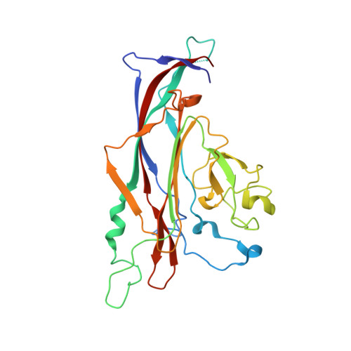
    Polyomaviruses are a family of ubiquitous double-stranded DNA viruses many of which are human pathogens. These include BK polyomavirus which causes severe urinary tract infection in immunocompromised patients and Merkel cell polyomavirus associated with aggressive cancers. The small genome of polyomaviruses lacks conventional drug targets, and no specific drugs are available at present. Here we focus on the main structural protein VP1 of BK polyomavirus which is responsible for icosahedral capsid formation. To provide a foundation towards rational drug design, we crystallized truncated VP1 pentamers and subjected them to a high-throughput screening for binding drug-like fragments through a direct X-ray analysis. To enable a highly performant screening, rigorous optimization of the crystallographic pipeline and processing with the latest generation PanDDA2 software were necessary. As a result, a total of 144 binding hits were established. Importantly, the hits are well clustered in six surface pockets. Three pockets are located on the outside of the pentamer and map on the regions where the 'invading' C-terminal arm of another pentamer is attached upon capsid assembly. Another set of three pockets is situated within the wide pore along the five-fold axis of the VP1 pentamer. These pockets are situated at the interaction interface with the minor capsid protein VP2 which is indispensable for normal functioning of the virus. Here we systematically analyse the three outside pockets which are highly conserved across various polyomaviruses, while point mutations in these pockets are detrimental for viral replication. We show that one of the pockets can accommodate antipsychotic drug trifluoperazine. For each pocket, we derive pharmacophore features which enable the design of small molecules preventing the interaction between VP1 pentamers and therefore inhibiting capsid assembly. Our data lay a foundation towards a rational development of first-in-class drugs targeting polyomavirus capsid.


  • Organizational Affiliation

    Biocrystallography, KU Leuven Herestraat 49 Leuven Belgium sergei.strelkov@kuleuven.be.


Macromolecules
Find similar proteins by:  (by identity cutoff)  |  3D Structure
Entity ID: 1
MoleculeChains Sequence LengthOrganismDetailsImage
Major capsid protein VP1A [auth AAA],
B [auth BBB],
C [auth CCC],
D [auth DDD],
E [auth EEE]
270Betapolyomavirus hominisMutation(s): 0 
UniProt
Find proteins for P03088 (BK polyomavirus)
Explore P03088 
Go to UniProtKB:  P03088
Entity Groups  
Sequence Clusters30% Identity50% Identity70% Identity90% Identity95% Identity100% Identity
UniProt GroupP03088
Sequence Annotations
Expand
  • Reference Sequence
Small Molecules
Ligands 4 Unique
IDChains Name / Formula / InChI Key2D Diagram3D Interactions
IOD
Query on IOD

Download Ideal Coordinates CCD File 
BA [auth CCC]
CA [auth CCC]
DA [auth CCC]
EA [auth CCC]
FA [auth CCC]
BA [auth CCC],
CA [auth CCC],
DA [auth CCC],
EA [auth CCC],
FA [auth CCC],
M [auth AAA],
MA [auth DDD],
N [auth AAA],
NA [auth DDD],
O [auth AAA],
OA [auth DDD],
TA [auth EEE],
U [auth BBB],
UA [auth EEE],
V [auth BBB],
VA [auth EEE]
IODIDE ION
I
XMBWDFGMSWQBCA-UHFFFAOYSA-M
DMS
Query on DMS

Download Ideal Coordinates CCD File 
AA [auth CCC]
JA [auth DDD]
K [auth AAA]
KA [auth DDD]
L [auth AAA]
AA [auth CCC],
JA [auth DDD],
K [auth AAA],
KA [auth DDD],
L [auth AAA],
LA [auth DDD],
RA [auth EEE],
S [auth BBB],
SA [auth EEE],
T [auth BBB],
Y [auth CCC],
Z [auth CCC]
DIMETHYL SULFOXIDE
C2 H6 O S
IAZDPXIOMUYVGZ-UHFFFAOYSA-N
CA
Query on CA

Download Ideal Coordinates CCD File 
F [auth AAA]
G [auth AAA]
GA [auth DDD]
H [auth AAA]
HA [auth DDD]
F [auth AAA],
G [auth AAA],
GA [auth DDD],
H [auth AAA],
HA [auth DDD],
I [auth AAA],
IA [auth DDD],
J [auth AAA],
Q [auth BBB],
QA [auth EEE],
R [auth BBB],
W [auth CCC],
X [auth CCC]
CALCIUM ION
Ca
BHPQYMZQTOCNFJ-UHFFFAOYSA-N
NA
Query on NA

Download Ideal Coordinates CCD File 
P [auth AAA],
PA [auth DDD]
SODIUM ION
Na
FKNQFGJONOIPTF-UHFFFAOYSA-N
Experimental Data & Validation

Experimental Data

  • Method: X-RAY DIFFRACTION
  • Resolution: 1.44 Å
  • R-Value Free: 0.186 
  • R-Value Work: 0.162 
  • Space Group: P 21 21 2
Unit Cell:
Length ( Å )Angle ( ˚ )
a = 138.622α = 90
b = 149.719β = 90
c = 65.488γ = 90
Software Package:
Software NamePurpose
REFMACrefinement
XDSdata reduction
Aimlessdata scaling
MOLREPphasing

Structure Validation

View Full Validation Report


Entry History & Funding Information

Deposition Data


Funding OrganizationLocationGrant Number
Research Foundation - Flanders (FWO)BelgiumG0A5718N

Revision History  (Full details and data files)

  • Version 1.0: 2022-06-22
    Type: Initial release
  • Version 1.1: 2022-08-31
    Changes: Database references, Derived calculations
  • Version 1.2: 2024-01-31
    Changes: Data collection, Refinement description