7B59

X-ray crystal structure of Sporosarcina pasteurii urease inhibited by Ag(PEt3)Br determined at 1.63 Angstroms


Experimental Data Snapshot

  • Method: X-RAY DIFFRACTION
  • Resolution: 1.63 Å
  • R-Value Free: 0.177 
  • R-Value Work: 0.149 

wwPDB Validation   3D Report Full Report


This is version 2.1 of the entry. See complete history


Literature

Kinetic and structural analysis of the inactivation of urease by mixed-ligand phosphine halide Ag(I) complexes.

Mazzei, L.Cirri, D.Cianci, M.Messori, L.Ciurli, S.

(2021) J Inorg Biochem 218: 111375-111375

  • DOI: https://doi.org/10.1016/j.jinorgbio.2021.111375
  • Primary Citation of Related Structures:  
    7B58, 7B59, 7B5A

  • PubMed Abstract: 

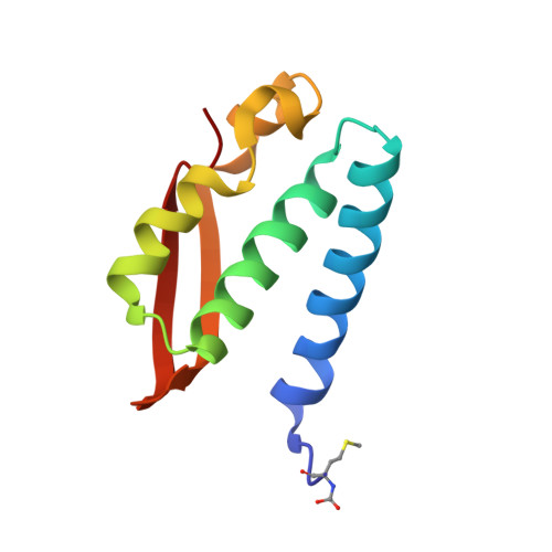
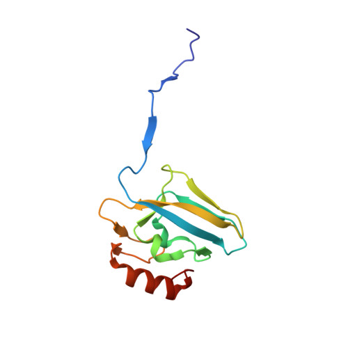
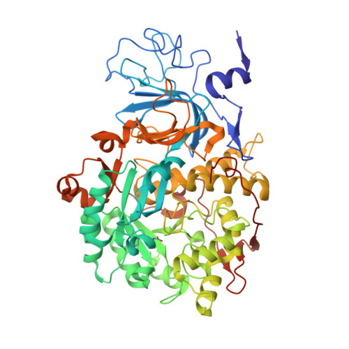
    Soft metal ions can inactivate urease, a Ni(II)-dependent enzyme whose hydrolytic activity has significant implications in agro-environmental science and human health. Kinetic and structural studies of the reaction of Canavalia ensiformis urease (JBU) and Sporosarcina pasteurii urease (SPU) with Ag(I) compounds of general formula [Ag(PEt 3 )X] 4 (X = Cl, Br, I), and with the ionic species [Ag(PEt 3 ) 2 ]NO 3 , revealed the role of the Ag(I) ion and its ligands in modulating the metal-enzyme interaction. The activity of JBU is obliterated by the [Ag(PEt 3 )X] 4 complexes, with IC 50 values in the nanomolar range; the efficiency of the inhibition increases in the Cl - < Br - < I - order. The activity of JBU upon [Ag(PEt 3 ) 2 ]NO 3 addition decreases to a plateau corresponding to ca. 60% of the original activity and decreases with time at a reduced rate. Synchrotron X-ray crystallography on single crystals obtained after the incubation of SPU with the Ag(I) complexes yielded high-resolution (1.63-1.97 Å) structures. The metal-protein adducts entail a dinuclear Ag(I) cluster bound to the conserved residues αCys322, αHis323, and αMet367, with a bridging cysteine thiolate atom, a weak Ag Ag bond, and a quasi-linear Ag(I) coordination geometry. These observations suggest a mechanism that involves the initial substitution of the phosphine ligand, followed by a structural rearrangement to yield the dinuclear Ag(I) cluster. These findings indicate that urease, in addition to the active site dinuclear Ni(II) cluster, possesses a secondary metal binding site, located on the mobile flap domain, capable of recognizing pairs of soft metal ions and controlling catalysis.


  • Organizational Affiliation

    Laboratory of Bioinorganic Chemistry, Department of Pharmacy and Biotechnology (FaBiT), University of Bologna, Via Giuseppe Fanin 40, I-40127 Bologna, Italy. Electronic address: luca.mazzei2@unibo.it.


Macromolecules
Find similar proteins by:  (by identity cutoff)  |  3D Structure
Entity ID: 1
MoleculeChains Sequence LengthOrganismDetailsImage
Urease subunit gammaA [auth AAA]100Sporosarcina pasteuriiMutation(s): 2 
EC: 3.5.1.5
UniProt
Find proteins for P41022 (Sporosarcina pasteurii)
Explore P41022 
Go to UniProtKB:  P41022
Entity Groups  
Sequence Clusters30% Identity50% Identity70% Identity90% Identity95% Identity100% Identity
UniProt GroupP41022
Sequence Annotations
Expand
  • Reference Sequence
Find similar proteins by:  (by identity cutoff)  |  3D Structure
Entity ID: 2
MoleculeChains Sequence LengthOrganismDetailsImage
Urease subunit betaB [auth BBB]122Sporosarcina pasteuriiMutation(s): 0 
EC: 3.5.1.5
UniProt
Find proteins for P41021 (Sporosarcina pasteurii)
Explore P41021 
Go to UniProtKB:  P41021
Entity Groups  
Sequence Clusters30% Identity50% Identity70% Identity90% Identity95% Identity100% Identity
UniProt GroupP41021
Sequence Annotations
Expand
  • Reference Sequence
Find similar proteins by:  (by identity cutoff)  |  3D Structure
Entity ID: 3
MoleculeChains Sequence LengthOrganismDetailsImage
Urease subunit alphaC [auth CCC]570Sporosarcina pasteuriiMutation(s): 0 
EC: 3.5.1.5
UniProt
Find proteins for P41020 (Sporosarcina pasteurii)
Explore P41020 
Go to UniProtKB:  P41020
Entity Groups  
Sequence Clusters30% Identity50% Identity70% Identity90% Identity95% Identity100% Identity
UniProt GroupP41020
Sequence Annotations
Expand
  • Reference Sequence
Small Molecules
Ligands 5 Unique
IDChains Name / Formula / InChI Key2D Diagram3D Interactions
AG (Subject of Investigation/LOI)
Query on AG

Download Ideal Coordinates CCD File 
X [auth CCC],
Y [auth CCC]
SILVER ION
Ag
FOIXSVOLVBLSDH-UHFFFAOYSA-N
SO4
Query on SO4

Download Ideal Coordinates CCD File 
BA [auth CCC]
CA [auth CCC]
DA [auth CCC]
EA [auth CCC]
FA [auth CCC]
BA [auth CCC],
CA [auth CCC],
DA [auth CCC],
EA [auth CCC],
FA [auth CCC],
GA [auth CCC],
H [auth AAA],
HA [auth CCC],
IA [auth CCC],
JA [auth CCC],
KA [auth CCC],
LA [auth CCC],
M [auth BBB],
MA [auth CCC],
N [auth BBB],
NA [auth CCC],
O [auth BBB],
P [auth BBB]
SULFATE ION
O4 S
QAOWNCQODCNURD-UHFFFAOYSA-L
EDO
Query on EDO

Download Ideal Coordinates CCD File 
AA [auth CCC]
D [auth AAA]
E [auth AAA]
F [auth AAA]
G [auth AAA]
AA [auth CCC],
D [auth AAA],
E [auth AAA],
F [auth AAA],
G [auth AAA],
I [auth BBB],
J [auth BBB],
K [auth BBB],
L [auth BBB],
S [auth CCC],
T [auth CCC],
V [auth CCC],
W [auth CCC],
Z [auth CCC]
1,2-ETHANEDIOL
C2 H6 O2
LYCAIKOWRPUZTN-UHFFFAOYSA-N
NI
Query on NI

Download Ideal Coordinates CCD File 
Q [auth CCC],
R [auth CCC]
NICKEL (II) ION
Ni
VEQPNABPJHWNSG-UHFFFAOYSA-N
O
Query on O

Download Ideal Coordinates CCD File 
U [auth CCC]OXYGEN ATOM
O
XLYOFNOQVPJJNP-UHFFFAOYSA-N
Modified Residues  2 Unique
IDChains TypeFormula2D DiagramParent
CXM
Query on CXM
A [auth AAA]L-PEPTIDE LINKINGC6 H11 N O4 SMET
KCX
Query on KCX
C [auth CCC]L-PEPTIDE LINKINGC7 H14 N2 O4LYS
Experimental Data & Validation

Experimental Data

  • Method: X-RAY DIFFRACTION
  • Resolution: 1.63 Å
  • R-Value Free: 0.177 
  • R-Value Work: 0.149 
  • Space Group: P 63 2 2
Unit Cell:
Length ( Å )Angle ( ˚ )
a = 131.41α = 90
b = 131.41β = 90
c = 189.416γ = 120
Software Package:
Software NamePurpose
REFMACrefinement
XDSdata reduction
Aimlessdata scaling
REFMACphasing

Structure Validation

View Full Validation Report


Entry History 

Deposition Data

Revision History  (Full details and data files)

  • Version 1.0: 2021-10-13
    Type: Initial release
  • Version 2.0: 2022-04-20
    Changes: Atomic model, Data collection, Database references, Source and taxonomy
  • Version 2.1: 2024-01-31
    Changes: Data collection, Derived calculations, Refinement description