7AUE

Melanocortin receptor 4 (MC4R) Gs protein complex


Experimental Data Snapshot

  • Method: ELECTRON MICROSCOPY
  • Resolution: 2.97 Å
  • Aggregation State: PARTICLE 
  • Reconstruction Method: SINGLE PARTICLE 

wwPDB Validation   3D Report Full Report


This is version 2.0 of the entry. See complete history


Literature

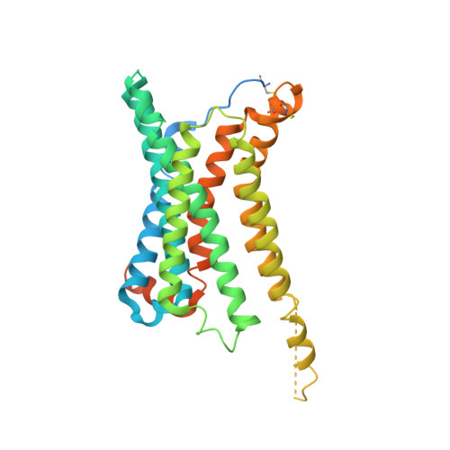
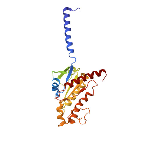
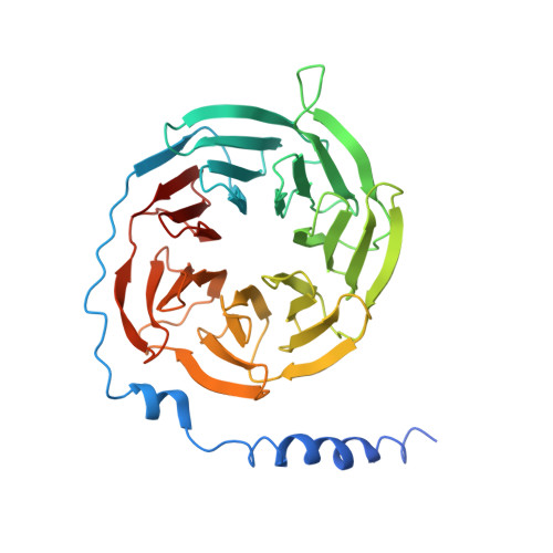
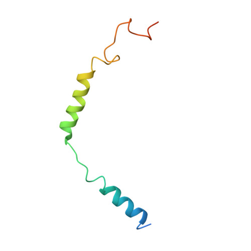
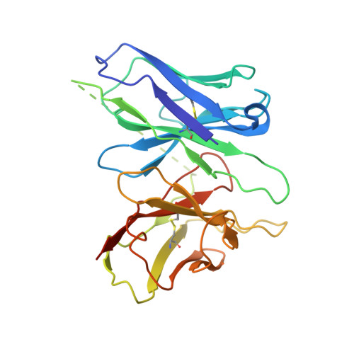
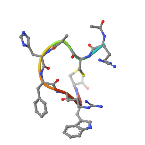
Structure reveals the activation mechanism of the MC4 receptor to initiate satiation signaling.

Israeli, H.Degtjarik, O.Fierro, F.Chunilal, V.Gill, A.K.Roth, N.J.Botta, J.Prabahar, V.Peleg, Y.Chan, L.F.Ben-Zvi, D.McCormick, P.J.Niv, M.Y.Shalev-Benami, M.

(2021) Science 372: 808-814

  • DOI: https://doi.org/10.1126/science.abf7958
  • Primary Citation of Related Structures:  
    7AUE

  • PubMed Abstract: 

    Obesity is a global epidemic that causes morbidity and impaired quality of life. The melanocortin receptor 4 (MC4R) is at the crux of appetite, energy homeostasis, and body-weight control in the central nervous system and is a prime target for anti-obesity drugs. Here, we present the cryo-electron microscopy (cryo-EM) structure of the human MC4R-G s signaling complex bound to the agonist setmelanotide, a cyclic peptide recently approved for the treatment of obesity. The work reveals the mechanism of MC4R activation, highlighting a molecular switch that initiates satiation signaling. In addition, our findings indicate that calcium (Ca 2+ ) is required for agonist, but not antagonist, efficacy. These results fill a gap in the understanding of MC4R activation and could guide the design of future weight-management drugs.


  • Organizational Affiliation

    Department of Chemical and Structural Biology, Weizmann Institute of Science, Rehovot 7610001, Israel.


Macromolecules
Find similar proteins by:  (by identity cutoff)  |  3D Structure
Entity ID: 1
MoleculeChains Sequence LengthOrganismDetailsImage
Melanocortin receptor 4A [auth R]350Homo sapiensMutation(s): 0 
Gene Names: MC4R
Membrane Entity: Yes 
UniProt & NIH Common Fund Data Resources
Find proteins for P32245 (Homo sapiens)
Explore P32245 
Go to UniProtKB:  P32245
PHAROS:  P32245
GTEx:  ENSG00000166603 
Entity Groups  
Sequence Clusters30% Identity50% Identity70% Identity90% Identity95% Identity100% Identity
UniProt GroupP32245
Sequence Annotations
Expand
  • Reference Sequence
Find similar proteins by:  (by identity cutoff)  |  3D Structure
Entity ID: 2
MoleculeChains Sequence LengthOrganismDetailsImage
Guanine nucleotide-binding protein G(s) subunit alpha isoforms shortB [auth A]373Homo sapiensMutation(s): 0 
Gene Names: GNASGNAS1GSP
Membrane Entity: Yes 
UniProt & NIH Common Fund Data Resources
Find proteins for P63092 (Homo sapiens)
Explore P63092 
Go to UniProtKB:  P63092
PHAROS:  P63092
GTEx:  ENSG00000087460 
Entity Groups  
Sequence Clusters30% Identity50% Identity70% Identity90% Identity95% Identity100% Identity
UniProt GroupP63092
Sequence Annotations
Expand
  • Reference Sequence
Find similar proteins by:  (by identity cutoff)  |  3D Structure
Entity ID: 3
MoleculeChains Sequence LengthOrganismDetailsImage
Guanine nucleotide-binding protein G(I)/G(S)/G(T) subunit beta-1C [auth B]357Homo sapiensMutation(s): 0 
Gene Names: GNB1
UniProt & NIH Common Fund Data Resources
Find proteins for P62873 (Homo sapiens)
Explore P62873 
Go to UniProtKB:  P62873
PHAROS:  P62873
GTEx:  ENSG00000078369 
Entity Groups  
Sequence Clusters30% Identity50% Identity70% Identity90% Identity95% Identity100% Identity
UniProt GroupP62873
Sequence Annotations
Expand
  • Reference Sequence
Find similar proteins by:  (by identity cutoff)  |  3D Structure
Entity ID: 4
MoleculeChains Sequence LengthOrganismDetailsImage
Guanine nucleotide-binding protein G(I)/G(S)/G(O) subunit gamma-2D [auth G]71Homo sapiensMutation(s): 0 
Gene Names: GNG2
Membrane Entity: Yes 
UniProt & NIH Common Fund Data Resources
Find proteins for P59768 (Homo sapiens)
Explore P59768 
Go to UniProtKB:  P59768
PHAROS:  P59768
GTEx:  ENSG00000186469 
Entity Groups  
Sequence Clusters30% Identity50% Identity70% Identity90% Identity95% Identity100% Identity
UniProt GroupP59768
Sequence Annotations
Expand
  • Reference Sequence
Find similar proteins by:  (by identity cutoff)  |  3D Structure
Entity ID: 5
MoleculeChains Sequence LengthOrganismDetailsImage
scFv16E [auth S]256Mus musculusMutation(s): 0 
Entity Groups  
Sequence Clusters30% Identity50% Identity70% Identity90% Identity95% Identity100% Identity
Sequence Annotations
Expand
  • Reference Sequence

Find similar proteins by:  Sequence   |   3D Structure  

Entity ID: 6
MoleculeChains Sequence LengthOrganismDetailsImage
AMINOSERINEF [auth C]9synthetic constructMutation(s): 0 
Sequence Annotations
Expand
  • Reference Sequence
Small Molecules
Ligands 1 Unique
IDChains Name / Formula / InChI Key2D Diagram3D Interactions
CA
Query on CA

Download Ideal Coordinates CCD File 
G [auth R]CALCIUM ION
Ca
BHPQYMZQTOCNFJ-UHFFFAOYSA-N
Modified Residues  1 Unique
IDChains TypeFormula2D DiagramParent
CY3
Query on CY3
F [auth C]L-PEPTIDE LINKINGC3 H8 N2 O SCYS
Experimental Data & Validation

Experimental Data

  • Method: ELECTRON MICROSCOPY
  • Resolution: 2.97 Å
  • Aggregation State: PARTICLE 
  • Reconstruction Method: SINGLE PARTICLE 

Structure Validation

View Full Validation Report


Entry History 

Deposition Data

Revision History  (Full details and data files)

  • Version 1.0: 2021-04-28
    Type: Initial release
  • Version 1.1: 2021-06-02
    Changes: Database references
  • Version 2.0: 2023-03-15
    Changes: Atomic model, Author supporting evidence, Data collection, Database references, Derived calculations, Non-polymer description, Polymer sequence, Source and taxonomy, Structure summary