7ASB

Crystal structure of dimeric chlorite dismutase variant Q74E (CCld Q74E) from Cyanothece sp. PCC7425


Experimental Data Snapshot

  • Method: X-RAY DIFFRACTION
  • Resolution: 1.40 Å
  • R-Value Free: 0.193 
  • R-Value Work: 0.162 
  • R-Value Observed: 0.162 

wwPDB Validation   3D Report Full Report


Ligand Structure Quality Assessment 


This is version 1.3 of the entry. See complete history


Literature

Arresting the Catalytic Arginine in Chlorite Dismutases: Impact on Heme Coordination, Thermal Stability, and Catalysis.

Schmidt, D.Serra, I.Mlynek, G.Pfanzagl, V.Hofbauer, S.Furtmuller, P.G.Djinovic-Carugo, K.Van Doorslaer, S.Obinger, C.

(2021) Biochemistry 60: 621-634

  • DOI: https://doi.org/10.1021/acs.biochem.0c00910
  • Primary Citation of Related Structures:  
    7ASB, 7ATI

  • PubMed Abstract: 

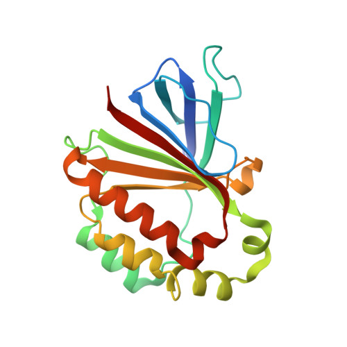
    Chlorite dismutases (Clds) are heme b -containing oxidoreductases that can decompose chlorite to chloride and molecular oxygen. They are divided in two clades that differ in oligomerization, subunit architecture, and the hydrogen-bonding network of the distal catalytic arginine, which is proposed to switch between two conformations during turnover. To understand the impact of the conformational dynamics of this basic amino acid on heme coordination, structure, and catalysis, Cld from Cyanothece sp. PCC7425 was used as a model enzyme. As typical for a clade 2 Cld, its distal arginine 127 is hydrogen-bonded to glutamine 74. The latter has been exchanged with either glutamate (Q74E) to arrest R127 in a salt bridge or valine (Q74V) that mirrors the setting in clade 1 Clds. We present the X-ray crystal structures of Q74V and Q74E and demonstrate the pH-induced changes in the environment and coordination of the heme iron by ultraviolet-visible, circular dichroism, and electron paramagnetic resonance spectroscopies as well as differential scanning calorimetry. The conformational dynamics of R127 is shown to have a significant role in heme coordination during the alkaline transition and in the thermal stability of the heme cavity, whereas its impact on the catalytic efficiency of chlorite degradation is relatively small. The findings are discussed with respect to (i) the flexible loop connecting the N-terminal and C-terminal ferredoxin-like domains, which differs in clade 1 and clade 2 Clds and carries Q74 in clade 2 proteins, and (ii) the proposed role(s) of the arginine in catalysis.


  • Organizational Affiliation

    Department of Chemistry, Institute of Biochemistry, University of Natural Resources and Life Sciences, Vienna, Muthgasse 18, A-1190 Vienna, Austria.


Macromolecules
Find similar proteins by:  (by identity cutoff)  |  3D Structure
Entity ID: 1
MoleculeChains Sequence LengthOrganismDetailsImage
Chlorite dismutase
A, B
188Cyanothece sp. PCC 7425Mutation(s): 1 
Gene Names: Cyan7425_1434
UniProt
Find proteins for B8HNS6 (Cyanothece sp. (strain PCC 7425 / ATCC 29141))
Explore B8HNS6 
Go to UniProtKB:  B8HNS6
Entity Groups  
Sequence Clusters30% Identity50% Identity70% Identity90% Identity95% Identity100% Identity
UniProt GroupB8HNS6
Sequence Annotations
Expand
  • Reference Sequence
Small Molecules
Ligands 5 Unique
IDChains Name / Formula / InChI Key2D Diagram3D Interactions
HEM (Subject of Investigation/LOI)
Query on HEM

Download Ideal Coordinates CCD File 
F [auth A],
N [auth B]
PROTOPORPHYRIN IX CONTAINING FE
C34 H32 Fe N4 O4
KABFMIBPWCXCRK-RGGAHWMASA-L
MES
Query on MES

Download Ideal Coordinates CCD File 
E [auth A]2-(N-MORPHOLINO)-ETHANESULFONIC ACID
C6 H13 N O4 S
SXGZJKUKBWWHRA-UHFFFAOYSA-N
PEG
Query on PEG

Download Ideal Coordinates CCD File 
I [auth A],
K [auth A],
L [auth B],
P [auth B]
DI(HYDROXYETHYL)ETHER
C4 H10 O3
MTHSVFCYNBDYFN-UHFFFAOYSA-N
SO4
Query on SO4

Download Ideal Coordinates CCD File 
J [auth A]SULFATE ION
O4 S
QAOWNCQODCNURD-UHFFFAOYSA-L
GOL
Query on GOL

Download Ideal Coordinates CCD File 
C [auth A]
D [auth A]
G [auth A]
H [auth A]
M [auth B]
C [auth A],
D [auth A],
G [auth A],
H [auth A],
M [auth B],
O [auth B]
GLYCEROL
C3 H8 O3
PEDCQBHIVMGVHV-UHFFFAOYSA-N
Experimental Data & Validation

Experimental Data

  • Method: X-RAY DIFFRACTION
  • Resolution: 1.40 Å
  • R-Value Free: 0.193 
  • R-Value Work: 0.162 
  • R-Value Observed: 0.162 
  • Space Group: P 1
Unit Cell:
Length ( Å )Angle ( ˚ )
a = 51.16α = 107.41
b = 52.738β = 99.143
c = 54.772γ = 108.984
Software Package:
Software NamePurpose
PHENIXrefinement
XDSdata reduction
XSCALEdata scaling
PHASERphasing

Structure Validation

View Full Validation Report


Ligand Structure Quality Assessment 


Entry History 

Deposition Data

Revision History  (Full details and data files)

  • Version 1.0: 2021-02-17
    Type: Initial release
  • Version 1.1: 2021-02-24
    Changes: Database references
  • Version 1.2: 2021-03-10
    Changes: Database references
  • Version 1.3: 2024-01-31
    Changes: Data collection, Database references, Refinement description