7API

THE S VARIANT OF HUMAN ALPHA1-ANTITRYPSIN, STRUCTURE AND IMPLICATIONS FOR FUNCTION AND METABOLISM


Experimental Data Snapshot

  • Method: X-RAY DIFFRACTION
  • Resolution: 3.00 Å
  • R-Value Work: 0.193 

wwPDB Validation   3D Report Full Report


This is version 2.1 of the entry. See complete history


Literature

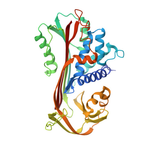
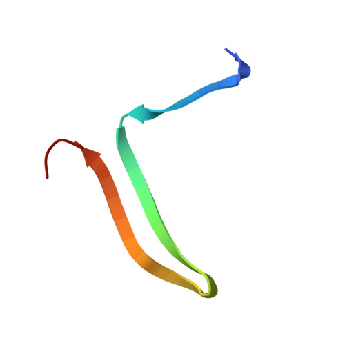
The S variant of human alpha 1-antitrypsin, structure and implications for function and metabolism.

Engh, R.Lobermann, H.Schneider, M.Wiegand, G.Huber, R.Laurell, C.B.

(1989) Protein Eng 2: 407-415

  • DOI: https://doi.org/10.1093/protein/2.6.407
  • Primary Citation of Related Structures:  
    7API, 8API, 9API

  • PubMed Abstract: 

    The S variant of the human alpha 1-antitrypsin with E-264----V, is responsible for a mild alpha 1-antitrypsin deficiency quite common in the European population. S protein specifically cleaved at the susceptible peptide bond was crystallized and its crystal structure determined and refined to 3.1 A resolution. The S variant crystallizes isomorphous to the normal M variant. The difference Fourier electron density map shows the E----V change as outstanding residual density. In addition, small structural changes of the main polypeptide chain radiate from the site of mutation and affect parts far removed from it. By the mutation, internal hydrogen bonds and salt linkages of E-264 to Y-38 and K-487, respectively, are lost. They cause the far-reaching slight distortions and are probably related to the reduced thermal stability of the S mutant. They may also be responsible for slower folding of the polypeptide chain and the clinical symptoms of alpha 1-antitrypsin deficiency. In a theoretical study by molecular dynamics methods simulations of the M and S proteins were made and the results analysed with respect to structural and dynamic properties and compared with the experimental results. There is a significant correlation between experimental and theoretical results in some respects.


  • Organizational Affiliation

    Max-Planck-Institut für Biochemie, Martinsried, FRG.


Macromolecules
Find similar proteins by:  (by identity cutoff)  |  3D Structure
Entity ID: 1
MoleculeChains Sequence LengthOrganismDetailsImage
ALPHA 1-ANTITRYPSIN347Homo sapiensMutation(s): 0 
UniProt & NIH Common Fund Data Resources
Find proteins for P01009 (Homo sapiens)
Explore P01009 
Go to UniProtKB:  P01009
PHAROS:  P01009
GTEx:  ENSG00000197249 
Entity Groups  
Sequence Clusters30% Identity50% Identity70% Identity90% Identity95% Identity100% Identity
UniProt GroupP01009
Sequence Annotations
Expand
  • Reference Sequence
Find similar proteins by:  (by identity cutoff)  |  3D Structure
Entity ID: 2
MoleculeChains Sequence LengthOrganismDetailsImage
ALPHA 1-ANTITRYPSIN36Homo sapiensMutation(s): 0 
UniProt & NIH Common Fund Data Resources
Find proteins for P01009 (Homo sapiens)
Explore P01009 
Go to UniProtKB:  P01009
PHAROS:  P01009
GTEx:  ENSG00000197249 
Entity Groups  
Sequence Clusters30% Identity50% Identity70% Identity90% Identity95% Identity100% Identity
UniProt GroupP01009
Sequence Annotations
Expand
  • Reference Sequence
Oligosaccharides

Help

Entity ID: 3
MoleculeChains Length2D Diagram Glycosylation3D Interactions
2-acetamido-2-deoxy-beta-D-glucopyranose-(1-2)-alpha-D-mannopyranose-(1-6)-[alpha-D-mannopyranose-(1-3)]alpha-D-mannopyranose-(1-4)-2-acetamido-2-deoxy-beta-D-glucopyranose-(1-4)-2-acetamido-2-deoxy-beta-D-glucopyranose
C
6N-Glycosylation
Glycosylation Resources
GlyTouCan:  G73276UD
GlyCosmos:  G73276UD
GlyGen:  G73276UD
Entity ID: 4
MoleculeChains Length2D Diagram Glycosylation3D Interactions
2-acetamido-2-deoxy-beta-D-glucopyranose-(1-4)-2-acetamido-2-deoxy-beta-D-glucopyranose
D, E
2N-Glycosylation
Glycosylation Resources
GlyTouCan:  G42666HT
GlyCosmos:  G42666HT
GlyGen:  G42666HT
Small Molecules
Ligands 1 Unique
IDChains Name / Formula / InChI Key2D Diagram3D Interactions
CYS
Query on CYS

Download Ideal Coordinates CCD File 
F [auth A]CYSTEINE
C3 H7 N O2 S
XUJNEKJLAYXESH-REOHCLBHSA-N
Experimental Data & Validation

Experimental Data

  • Method: X-RAY DIFFRACTION
  • Resolution: 3.00 Å
  • R-Value Work: 0.193 
  • Space Group: P 43 21 2
Unit Cell:
Length ( Å )Angle ( ˚ )
a = 120.5α = 90
b = 120.5β = 90
c = 113.5γ = 90
Software Package:
Software NamePurpose
EREFrefinement

Structure Validation

View Full Validation Report


Entry History 

Deposition Data

Revision History  (Full details and data files)

  • Version 1.0: 1990-10-15
    Type: Initial release
  • Version 1.1: 2008-03-25
    Changes: Version format compliance
  • Version 1.2: 2011-07-13
    Changes: Non-polymer description, Version format compliance
  • Version 2.0: 2020-07-29
    Type: Remediation
    Reason: Carbohydrate remediation
    Changes: Advisory, Atomic model, Data collection, Derived calculations, Other, Structure summary
  • Version 2.1: 2024-01-10
    Changes: Advisory, Data collection, Database references, Derived calculations, Structure summary