6ZUR

Psychrophilic aromatic amino acids aminotransferase from Psychrobacter sp. B6 cocrystalized with substrate analog - L-p-hydroxyphenyllactic acid


Experimental Data Snapshot

  • Method: X-RAY DIFFRACTION
  • Resolution: 2.31 Å
  • R-Value Free: 0.246 
  • R-Value Work: 0.198 
  • R-Value Observed: 0.200 

wwPDB Validation   3D Report Full Report


Ligand Structure Quality Assessment 


This is version 2.1 of the entry. See complete history


Literature

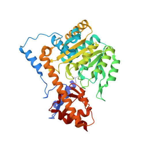
Structural Evidence of Active Site Adaptability towards Different Sized Substrates of Aromatic Amino Acid Aminotransferase from Psychrobacter Sp. B6.

Bujacz, A.Rum, J.Rutkiewicz, M.Pietrzyk-Brzezinska, A.J.Bujacz, G.

(2021) Materials (Basel) 14

  • DOI: https://doi.org/10.3390/ma14123351
  • Primary Citation of Related Structures:  
    6T3V, 6ZUP, 6ZUR, 6ZVG

  • PubMed Abstract: 

    Aromatic amino acid aminotransferases present a special potential in the production of drugs and synthons, thanks to their ability to accommodate a wider range of substrates in their active site, in contrast to aliphatic amino acid aminotransferases. The mechanism of active site adjustment toward substrates of psychrophilic aromatic amino acid aminotransferase ( Psy ArAT) from Psychrobacter sp. B6 is discussed based on crystal structures of complexes with four hydroxy-analogs of substrates: phenylalanine, tyrosine, tryptophan and aspartic acid. These competitive inhibitors are bound in the active center of Psy ArAT but do not undergo transamination reaction, which makes them an outstanding tool for examination of the enzyme catalytic center. The use of hydroxy-acids enabled insight into substrate binding by native Psy ArAT, without mutating the catalytic lysine and modifying cofactor interactions. Thus, the binding mode of substrates and the resulting analysis of the volume of the catalytic site is close to a native condition. Observation of these inhibitors' binding allows for explanation of the enzyme's adaptability to process various sizes of substrates and to gain knowledge about its potential biotechnological application. Depending on the character and size of the used inhibitors, the enzyme crystallized in different space groups and showed conformational changes of the active site upon ligand binding.


  • Organizational Affiliation

    Institute of Molecular and Industrial Biotechnology, Lodz University of Technology, Stefanowskiego 4/10, 90-924 Lodz, Poland.


Macromolecules
Find similar proteins by:  (by identity cutoff)  |  3D Structure
Entity ID: 1
MoleculeChains Sequence LengthOrganismDetailsImage
Aminotransferase
A, B
398Psychrobacter sp. B6Mutation(s): 0 
EC: 2.6.1
UniProt
Find proteins for C7E5X4 (Psychrobacter sp. B6)
Explore C7E5X4 
Go to UniProtKB:  C7E5X4
Entity Groups  
Sequence Clusters30% Identity50% Identity70% Identity90% Identity95% Identity100% Identity
UniProt GroupC7E5X4
Sequence Annotations
Expand
  • Reference Sequence
Small Molecules
Ligands 4 Unique
IDChains Name / Formula / InChI Key2D Diagram3D Interactions
PLP (Subject of Investigation/LOI)
Query on PLP

Download Ideal Coordinates CCD File 
L [auth A],
Q [auth B]
PYRIDOXAL-5'-PHOSPHATE
C8 H10 N O6 P
NGVDGCNFYWLIFO-UHFFFAOYSA-N
TYF (Subject of Investigation/LOI)
Query on TYF

Download Ideal Coordinates CCD File 
E [auth A],
F [auth A]
(2S)-2-hydroxy-3-(4-hydroxyphenyl)propanoic acid
C9 H10 O4
JVGVDSSUAVXRDY-QMMMGPOBSA-N
NO3
Query on NO3

Download Ideal Coordinates CCD File 
G [auth A]
H [auth A]
I [auth A]
J [auth A]
K [auth A]
G [auth A],
H [auth A],
I [auth A],
J [auth A],
K [auth A],
M [auth B],
N [auth B],
O [auth B],
P [auth B]
NITRATE ION
N O3
NHNBFGGVMKEFGY-UHFFFAOYSA-N
MG
Query on MG

Download Ideal Coordinates CCD File 
C [auth A],
D [auth A]
MAGNESIUM ION
Mg
JLVVSXFLKOJNIY-UHFFFAOYSA-N
Experimental Data & Validation

Experimental Data

  • Method: X-RAY DIFFRACTION
  • Resolution: 2.31 Å
  • R-Value Free: 0.246 
  • R-Value Work: 0.198 
  • R-Value Observed: 0.200 
  • Space Group: P 1 21 1
Unit Cell:
Length ( Å )Angle ( ˚ )
a = 71.73α = 90
b = 63.858β = 102.67
c = 82.847γ = 90
Software Package:
Software NamePurpose
REFMACrefinement
XDSdata reduction
XSCALEdata scaling
MOLREPphasing
PDB_EXTRACTdata extraction

Structure Validation

View Full Validation Report


Ligand Structure Quality Assessment 


Entry History & Funding Information

Deposition Data


Funding OrganizationLocationGrant Number
Polish National Science CentrePoland2016/21/B/ST5/00555

Revision History  (Full details and data files)

  • Version 1.0: 2021-07-14
    Type: Initial release
  • Version 2.0: 2023-11-15
    Changes: Atomic model, Data collection, Database references
  • Version 2.1: 2024-01-31
    Changes: Refinement description