6ZSW

M2 mutant (R111K:Y134F:T54V:R132Q:P39Y:R59Y) of human cellular retinoic acid binding protein II - 6 conjugate


Experimental Data Snapshot

  • Method: X-RAY DIFFRACTION
  • Resolution: 2.08 Å
  • R-Value Free: 0.228 
  • R-Value Work: 0.207 
  • R-Value Observed: 0.208 

wwPDB Validation   3D Report Full Report


Ligand Structure Quality Assessment 


This is version 1.3 of the entry. See complete history


Literature

Xanthopsin-Like Systems via Site-Specific Click-Functionalization of a Retinoic Acid Binding Protein.

Tassone, G.Paolino, M.Pozzi, C.Reale, A.Salvini, L.Giorgi, G.Orlandini, M.Galvagni, F.Mangani, S.Yang, X.Carlotti, B.Ortica, F.Latterini, L.Olivucci, M.Cappelli, A.

(2022) Chembiochem 23: e202100449-e202100449

  • DOI: https://doi.org/10.1002/cbic.202100449
  • Primary Citation of Related Structures:  
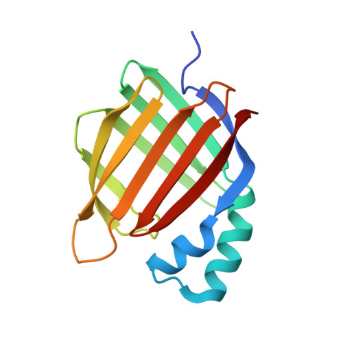
    6Z2U, 6Z2Z, 6ZSW, 6ZSX

  • PubMed Abstract: 

    The use of light-responsive proteins to control both living or synthetic cells, is at the core of the expanding fields of optogenetics and synthetic biology. It is thus apparent that a richer reaction toolbox for the preparation of such systems is of fundamental importance. Here, we provide a proof-of-principle demonstration that Morita-Baylis-Hillman adducts can be employed to perform a facile site-specific, irreversible and diastereoselective click-functionalization of a lysine residue buried into a lipophilic binding pocket and yielding an unnatural chromophore with an extended π-system. In doing so we effectively open the path to the in vitro preparation of a library of synthetic proteins structurally reminiscent of xanthopsin eubacterial photoreceptors. We argue that such a library, made of variable unnatural chromophores inserted in an easy-to-mutate and crystallize retinoic acid transporter, significantly expand the scope of the recently introduced rhodopsin mimics as both optogenetic and "lab-on-a-molecule" tools.


  • Organizational Affiliation

    Dipartimento di Biotecnologie, Chimica e Farmacia, Dipartimento di Eccellenza 2018-2022), Università degli Studi di Siena, Via A. Moro 2, 53100, Siena, Italy.


Macromolecules
Find similar proteins by:  (by identity cutoff)  |  3D Structure
Entity ID: 1
MoleculeChains Sequence LengthOrganismDetailsImage
Cellular retinoic acid-binding protein 2141Homo sapiensMutation(s): 6 
Gene Names: CRABP2
UniProt & NIH Common Fund Data Resources
Find proteins for P29373 (Homo sapiens)
Explore P29373 
Go to UniProtKB:  P29373
PHAROS:  P29373
GTEx:  ENSG00000143320 
Entity Groups  
Sequence Clusters30% Identity50% Identity70% Identity90% Identity95% Identity100% Identity
UniProt GroupP29373
Sequence Annotations
Expand
  • Reference Sequence
Small Molecules
Ligands 2 Unique
IDChains Name / Formula / InChI Key2D Diagram3D Interactions
QPB (Subject of Investigation/LOI)
Query on QPB

Download Ideal Coordinates CCD File 
B [auth A]methyl (~{Z})-3-(4-hydroxyphenyl)-2-methyl-prop-2-enoate
C11 H12 O3
CPKCCIUDOPJZSI-FPLPWBNLSA-N
ACT
Query on ACT

Download Ideal Coordinates CCD File 
C [auth A]ACETATE ION
C2 H3 O2
QTBSBXVTEAMEQO-UHFFFAOYSA-M
Experimental Data & Validation

Experimental Data

  • Method: X-RAY DIFFRACTION
  • Resolution: 2.08 Å
  • R-Value Free: 0.228 
  • R-Value Work: 0.207 
  • R-Value Observed: 0.208 
  • Space Group: P 31 2 1
Unit Cell:
Length ( Å )Angle ( ˚ )
a = 58.192α = 90
b = 58.192β = 90
c = 102.25γ = 120
Software Package:
Software NamePurpose
REFMACrefinement
PDB_EXTRACTdata extraction
XDSdata reduction
SCALAdata scaling
MOLREPphasing

Structure Validation

View Full Validation Report


Ligand Structure Quality Assessment 


Entry History 

Deposition Data

Revision History  (Full details and data files)

  • Version 1.0: 2021-07-28
    Type: Initial release
  • Version 1.1: 2021-11-17
    Changes: Data collection, Database references
  • Version 1.2: 2022-01-19
    Changes: Data collection, Database references
  • Version 1.3: 2024-01-31
    Changes: Data collection, Refinement description