6ZS5

3.5 A cryo-EM structure of human uromodulin filament core


Experimental Data Snapshot

  • Method: ELECTRON MICROSCOPY
  • Resolution: 3.50 Å
  • Aggregation State: FILAMENT 
  • Reconstruction Method: SINGLE PARTICLE 

wwPDB Validation   3D Report Full Report


This is version 1.0 of the entry. See complete history


Literature

The cryo-EM structure of the human uromodulin filament core reveals a unique assembly mechanism.

Stanisich, J.J.Zyla, D.S.Afanasyev, P.Xu, J.Kipp, A.Olinger, E.Devuyst, O.Pilhofer, M.Boehringer, D.Glockshuber, R.

(2020) Elife 9

  • DOI: https://doi.org/10.7554/eLife.60265
  • Primary Citation of Related Structures:  
    6ZS5, 6ZYA

  • PubMed Abstract: 

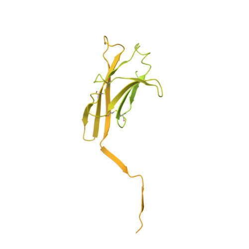
    The glycoprotein uromodulin (UMOD) is the most abundant protein in human urine and forms filamentous homopolymers that encapsulate and aggregate uropathogens, promoting pathogen clearance by urine excretion. Despite its critical role in the innate immune response against urinary tract infections, the structural basis and mechanism of UMOD polymerization remained unknown. Here, we present the cryo-EM structure of the UMOD filament core at 3.5 Å resolution, comprised of the bipartite zona pellucida (ZP) module in a helical arrangement with a rise of ~65 Å and a twist of ~180°. The immunoglobulin-like ZPN and ZPC subdomains of each monomer are separated by a long linker that interacts with the preceding ZPC and following ZPN subdomains by β-sheet complementation. The unique filament architecture suggests an assembly mechanism in which subunit incorporation could be synchronized with proteolytic cleavage of the C-terminal pro-peptide that anchors assembly-incompetent UMOD precursors to the membrane.


  • Organizational Affiliation

    Institute of Molecular Biology & Biophysics, ETH Zurich, Zurich, Switzerland.


Macromolecules
Find similar proteins by:  (by identity cutoff)  |  3D Structure
Entity ID: 1
MoleculeChains Sequence LengthOrganismDetailsImage
UromodulinA,
B [auth D]
640Homo sapiensMutation(s): 0 
UniProt & NIH Common Fund Data Resources
Find proteins for P07911 (Homo sapiens)
Explore P07911 
Go to UniProtKB:  P07911
PHAROS:  P07911
GTEx:  ENSG00000169344 
Entity Groups  
Sequence Clusters30% Identity50% Identity70% Identity90% Identity95% Identity100% Identity
UniProt GroupP07911
Sequence Annotations
Expand
  • Reference Sequence
Oligosaccharides

Help

Entity ID: 2
MoleculeChains Length2D Diagram Glycosylation3D Interactions
alpha-D-mannopyranose-(1-3)-[alpha-D-mannopyranose-(1-6)]beta-D-mannopyranose-(1-4)-2-acetamido-2-deoxy-beta-D-glucopyranose-(1-4)-2-acetamido-2-deoxy-beta-D-glucopyranoseC [auth B]5N-Glycosylation
Glycosylation Resources
GlyTouCan:  G22768VO
GlyCosmos:  G22768VO
GlyGen:  G22768VO
Entity ID: 3
MoleculeChains Length2D Diagram Glycosylation3D Interactions
2-acetamido-2-deoxy-beta-D-glucopyranose-(1-4)-2-acetamido-2-deoxy-beta-D-glucopyranoseD [auth C]2N-Glycosylation
Glycosylation Resources
GlyTouCan:  G42666HT
GlyCosmos:  G42666HT
GlyGen:  G42666HT
Experimental Data & Validation

Experimental Data

  • Method: ELECTRON MICROSCOPY
  • Resolution: 3.50 Å
  • Aggregation State: FILAMENT 
  • Reconstruction Method: SINGLE PARTICLE 
EM Software:
TaskSoftware PackageVersion
MODEL REFINEMENTPHENIX1.18.2
MODEL REFINEMENTRosetta
RECONSTRUCTIONcryoSPARC2.15

Structure Validation

View Full Validation Report


Entry History & Funding Information

Deposition Data


Funding OrganizationLocationGrant Number
Swiss National Science FoundationSwitzerland310030B_176403/1
Swiss National Science FoundationSwitzerland31003A_156304
Swiss National Science FoundationSwitzerland31003A_179255
European Research Council (ERC)Switzerland679209

Revision History  (Full details and data files)

  • Version 1.0: 2020-09-02
    Type: Initial release