6ZQQ

Structure of the Pmt3-MIR domain with bound ligands


Experimental Data Snapshot

  • Method: X-RAY DIFFRACTION
  • Resolution: 1.90 Å
  • R-Value Free: 0.220 
  • R-Value Work: 0.174 
  • R-Value Observed: 0.176 

wwPDB Validation   3D Report Full Report


Ligand Structure Quality Assessment 


This is version 1.2 of the entry. See complete history


Literature

Functional implications of MIR domains in protein O -mannosylation.

Chiapparino, A.Grbavac, A.Jonker, H.R.Hackmann, Y.Mortensen, S.Zatorska, E.Schott, A.Stier, G.Saxena, K.Wild, K.Schwalbe, H.Strahl, S.Sinning, I.

(2020) Elife 9

  • DOI: https://doi.org/10.7554/eLife.61189
  • Primary Citation of Related Structures:  
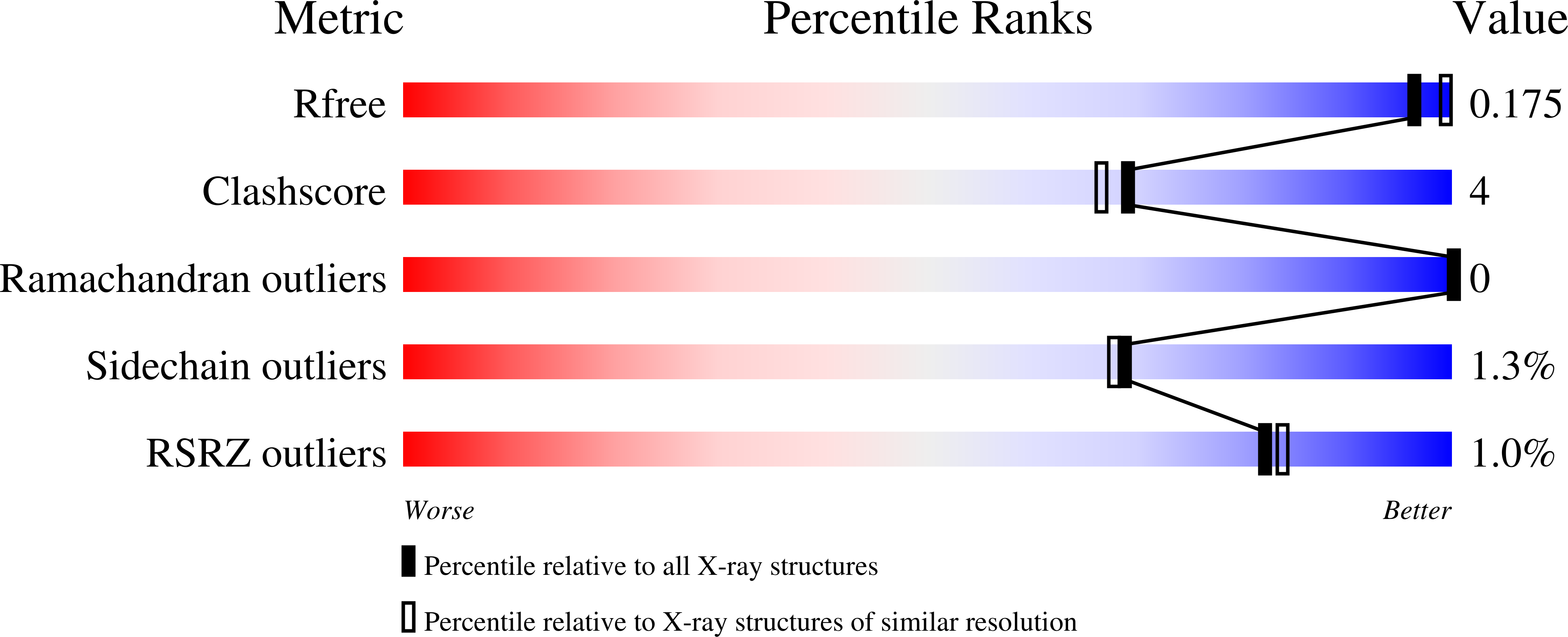
    6ZQP, 6ZQQ

  • PubMed Abstract: 

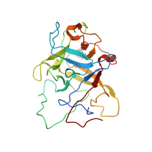
    Protein O -mannosyltransferases (PMTs) represent a conserved family of multispanning endoplasmic reticulum membrane proteins involved in glycosylation of S/T-rich protein substrates and unfolded proteins. PMTs work as dimers and contain a luminal MIR domain with a β-trefoil fold, which is susceptive for missense mutations causing α-dystroglycanopathies in humans. Here, we analyze PMT-MIR domains by an integrated structural biology approach using X-ray crystallography and NMR spectroscopy and evaluate their role in PMT function in vivo. We determine Pmt2- and Pmt3-MIR domain structures and identify two conserved mannose-binding sites, which are consistent with general β-trefoil carbohydrate-binding sites (α, β), and also a unique PMT2-subfamily exposed FKR motif. We show that conserved residues in site α influence enzyme processivity of the Pmt1-Pmt2 heterodimer in vivo. Integration of the data into the context of a Pmt1-Pmt2 structure and comparison with homologous β-trefoil - carbohydrate complexes allows for a functional description of MIR domains in protein O -mannosylation.


  • Organizational Affiliation

    Heidelberg University Biochemistry Center (BZH), Heidelberg, Germany.


Macromolecules
Find similar proteins by:  (by identity cutoff)  |  3D Structure
Entity ID: 1
MoleculeChains Sequence LengthOrganismDetailsImage
PMT3 isoform 1
A, B, C, D
212Saccharomyces cerevisiaeMutation(s): 0 
Gene Names: PMT3GI526_G0005657
Membrane Entity: Yes 
UniProt
Find proteins for P47190 (Saccharomyces cerevisiae (strain ATCC 204508 / S288c))
Explore P47190 
Go to UniProtKB:  P47190
Entity Groups  
Sequence Clusters30% Identity50% Identity70% Identity90% Identity95% Identity100% Identity
UniProt GroupP47190
Sequence Annotations
Expand
  • Reference Sequence
Experimental Data & Validation

Experimental Data

  • Method: X-RAY DIFFRACTION
  • Resolution: 1.90 Å
  • R-Value Free: 0.220 
  • R-Value Work: 0.174 
  • R-Value Observed: 0.176 
  • Space Group: P 1
Unit Cell:
Length ( Å )Angle ( ˚ )
a = 55.289α = 107.9
b = 64.306β = 99.85
c = 65.555γ = 99.72
Software Package:
Software NamePurpose
PHENIXrefinement
PDB_EXTRACTdata extraction
XDSdata reduction
Aimlessdata scaling
PHASERphasing

Structure Validation

View Full Validation Report


Ligand Structure Quality Assessment 


Entry History & Funding Information

Deposition Data


Funding OrganizationLocationGrant Number
German Research Foundation (DFG)--

Revision History  (Full details and data files)

  • Version 1.0: 2020-12-23
    Type: Initial release
  • Version 1.1: 2021-01-13
    Changes: Database references
  • Version 1.2: 2024-01-31
    Changes: Data collection, Database references, Refinement description