


Cryo-EM as a tool to study KatG from Mycobacterium tuberculosis
Blundell, T.L., Chaplin, A.K., Munir, A.To be published.
Experimental Data Snapshot
wwPDB Validation 3D Report Full Report

Entity ID: 1 | |||||
|---|---|---|---|---|---|
| Molecule | Chains | Sequence Length | Organism | Details | Image |
| Catalase-peroxidase | A [auth AP1], B [auth BP1] | 740 | Mycobacterium tuberculosis | Mutation(s): 0 Gene Names: katG, AYJ03_010035, ERS007661_00994, ERS013471_02729, ERS024276_01596, ERS075361_01376, ERS094182_01139, F6W99_00474, FRD82_12135 EC: 1.11.1.21 | 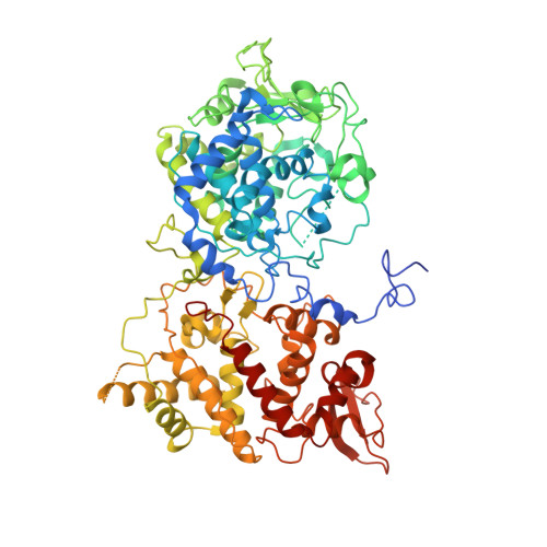 |
UniProt | |||||
Find proteins for P9WIE5 (Mycobacterium tuberculosis (strain ATCC 25618 / H37Rv)) Explore P9WIE5 Go to UniProtKB: P9WIE5 | |||||
Entity Groups | |||||
| Sequence Clusters | 30% Identity50% Identity70% Identity90% Identity95% Identity100% Identity | ||||
| UniProt Group | P9WIE5 | ||||
Sequence AnnotationsExpand | |||||
| |||||
| Ligands 1 Unique | |||||
|---|---|---|---|---|---|
| ID | Chains | Name / Formula / InChI Key | 2D Diagram | 3D Interactions | |
| HEM (Subject of Investigation/LOI) Query on HEM | C [auth AP1], D [auth BP1] | PROTOPORPHYRIN IX CONTAINING FE C34 H32 Fe N4 O4 KABFMIBPWCXCRK-RGGAHWMASA-L |  | ||
| Task | Software Package | Version |
|---|---|---|
| RECONSTRUCTION | cryoSPARC | |
| Funding Organization | Location | Grant Number |
|---|---|---|
| Bill & Melinda Gates Foundation | United Kingdom | RG 86546 |
RCSB PDB (citation) is hosted by
RCSB PDB is a member of the

