6ZHZ

OleP-oleandolide(DEO) in high salt crystallization conditions


Experimental Data Snapshot

  • Method: X-RAY DIFFRACTION
  • Resolution: 2.20 Å
  • R-Value Free: 0.243 
  • R-Value Work: 0.187 
  • R-Value Observed: 0.190 

wwPDB Validation   3D Report Full Report


Ligand Structure Quality Assessment 


This is version 1.1 of the entry. See complete history


Literature

Dissecting the Cytochrome P450 OleP Substrate Specificity: Evidence for a Preferential Substrate.

Parisi, G.Freda, I.Exertier, C.Cecchetti, C.Gugole, E.Cerutti, G.D'Auria, L.Macone, A.Vallone, B.Savino, C.Montemiglio, L.C.

(2020) Biomolecules 10

  • DOI: https://doi.org/10.3390/biom10101411
  • Primary Citation of Related Structures:  
    6ZHZ, 6ZI2, 6ZI3, 6ZI7

  • PubMed Abstract: 

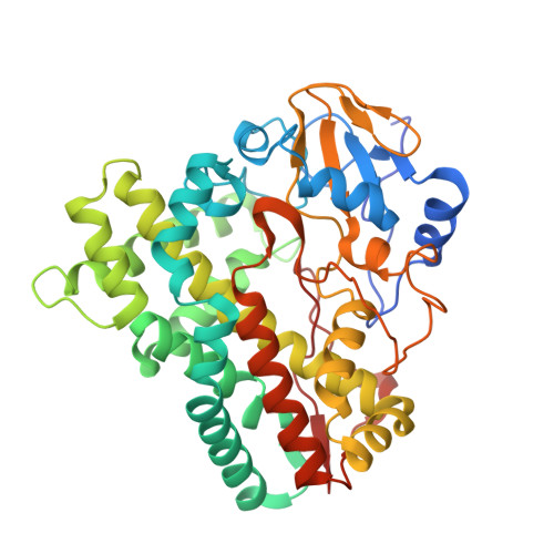
    The cytochrome P450 OleP catalyzes the epoxidation of aliphatic carbons on both the aglycone 8.8a-deoxyoleandolide (DEO) and the monoglycosylated L-olivosyl-8.8a-deoxyoleandolide (L-O-DEO) intermediates of oleandomycin biosynthesis. We investigated the substrate versatility of the enzyme. X-ray and equilibrium binding data show that the aglycone DEO loosely fits the OleP active site, triggering the closure that prepares it for catalysis only on a minor population of enzyme. The open-to-closed state transition allows solvent molecules to accumulate in a cavity that forms upon closure, mediating protein-substrate interactions. In silico docking of the monoglycosylated L-O-DEO in the closed OleP-DEO structure shows that the L-olivosyl moiety can be hosted in the same cavity, replacing solvent molecules and directly contacting structural elements involved in the transition. X-ray structures of aglycone-bound OleP in the presence of L-rhamnose confirm the cavity as a potential site for sugar binding. All considered, we propose L-O-DEO as the optimal substrate of OleP, the L-olivosyl moiety possibly representing the molecular wedge that triggers a more efficient structural response upon substrate binding, favoring and stabilizing the enzyme closure before catalysis. OleP substrate versatility is supported by structural solvent molecules that compensate for the absence of a glycosyl unit when the aglycone is bound.


  • Organizational Affiliation

    Istituto Pasteur-Fondazione Cenci Bolognetti and Department of Biochemical Sciences "Alessandro Rossi Fanelli", Sapienza, University of Rome, P. le Aldo Moro, 5, 00185 Rome, Italy.


Macromolecules
Find similar proteins by:  (by identity cutoff)  |  3D Structure
Entity ID: 1
MoleculeChains Sequence LengthOrganismDetailsImage
Cytochrome P-450
A, B, C, D, E
A, B, C, D, E, F
407Streptomyces antibioticusMutation(s): 0 
Gene Names: oleP
UniProt
Find proteins for Q59819 (Streptomyces antibioticus)
Explore Q59819 
Go to UniProtKB:  Q59819
Entity Groups  
Sequence Clusters30% Identity50% Identity70% Identity90% Identity95% Identity100% Identity
UniProt GroupQ59819
Sequence Annotations
Expand
  • Reference Sequence
Small Molecules
Ligands 6 Unique
IDChains Name / Formula / InChI Key2D Diagram3D Interactions
HEM
Query on HEM

Download Ideal Coordinates CCD File 
G [auth A]
GB [auth C]
HD [auth F]
KC [auth D]
XC [auth E]
G [auth A],
GB [auth C],
HD [auth F],
KC [auth D],
XC [auth E],
Z [auth B]
PROTOPORPHYRIN IX CONTAINING FE
C34 H32 Fe N4 O4
KABFMIBPWCXCRK-RGGAHWMASA-L
QR8 (Subject of Investigation/LOI)
Query on QR8

Download Ideal Coordinates CCD File 
AA [auth B]
H [auth A]
HB [auth C]
ID [auth F]
LC [auth D]
AA [auth B],
H [auth A],
HB [auth C],
ID [auth F],
LC [auth D],
YC [auth E]
(3~{R},4~{S},5~{R},6~{S},7~{S},9~{S},11~{R},12~{S},13~{R},14~{R})-3,5,7,9,11,13,14-heptamethyl-4,6,12-tris(oxidanyl)-1-oxacyclotetradecane-2,10-dione
C20 H36 O6
OLPAVFVRSHWADO-ODNSKKOKSA-N
TRS
Query on TRS

Download Ideal Coordinates CCD File 
I [auth A],
JD [auth F],
MC [auth D]
2-AMINO-2-HYDROXYMETHYL-PROPANE-1,3-DIOL
C4 H12 N O3
LENZDBCJOHFCAS-UHFFFAOYSA-O
GOL
Query on GOL

Download Ideal Coordinates CCD File 
AB [auth B]
BB [auth B]
CB [auth B]
DB [auth B]
DC [auth C]
AB [auth B],
BB [auth B],
CB [auth B],
DB [auth B],
DC [auth C],
EB [auth B],
EC [auth C],
FC [auth C],
GC [auth C],
HC [auth C],
IC [auth C],
SD [auth F],
VC [auth D],
X [auth A],
ZA [auth B]
GLYCEROL
C3 H8 O3
PEDCQBHIVMGVHV-UHFFFAOYSA-N
FMT
Query on FMT

Download Ideal Coordinates CCD File 
AC [auth C]
AD [auth E]
BA [auth B]
BC [auth C]
BD [auth E]
AC [auth C],
AD [auth E],
BA [auth B],
BC [auth C],
BD [auth E],
CA [auth B],
CC [auth C],
CD [auth E],
DA [auth B],
DD [auth E],
EA [auth B],
ED [auth E],
FA [auth B],
FD [auth E],
GA [auth B],
GD [auth E],
HA [auth B],
IA [auth B],
IB [auth C],
J [auth A],
JA [auth B],
JB [auth C],
K [auth A],
KA [auth B],
KB [auth C],
KD [auth F],
L [auth A],
LA [auth B],
LB [auth C],
LD [auth F],
M [auth A],
MA [auth B],
MB [auth C],
MD [auth F],
N [auth A],
NA [auth B],
NB [auth C],
NC [auth D],
ND [auth F],
O [auth A],
OA [auth B],
OB [auth C],
OC [auth D],
OD [auth F],
P [auth A],
PA [auth B],
PB [auth C],
PC [auth D],
PD [auth F],
Q [auth A],
QA [auth B],
QB [auth C],
QC [auth D],
QD [auth F],
R [auth A],
RA [auth B],
RB [auth C],
RC [auth D],
RD [auth F],
S [auth A],
SA [auth B],
SB [auth C],
SC [auth D],
T [auth A],
TA [auth B],
TB [auth C],
TC [auth D],
U [auth A],
UA [auth B],
UB [auth C],
UC [auth D],
V [auth A],
VA [auth B],
VB [auth C],
W [auth A],
WA [auth B],
WB [auth C],
XA [auth B],
XB [auth C],
YA [auth B],
YB [auth C],
ZB [auth C],
ZC [auth E]
FORMIC ACID
C H2 O2
BDAGIHXWWSANSR-UHFFFAOYSA-N
NA
Query on NA

Download Ideal Coordinates CCD File 
FB [auth B],
JC [auth C],
TD [auth F],
WC [auth D],
Y [auth A]
SODIUM ION
Na
FKNQFGJONOIPTF-UHFFFAOYSA-N
Experimental Data & Validation

Experimental Data

  • Method: X-RAY DIFFRACTION
  • Resolution: 2.20 Å
  • R-Value Free: 0.243 
  • R-Value Work: 0.187 
  • R-Value Observed: 0.190 
  • Space Group: C 1 2 1
Unit Cell:
Length ( Å )Angle ( ˚ )
a = 247.469α = 90
b = 111.215β = 129.39
c = 159.203γ = 90
Software Package:
Software NamePurpose
REFMACrefinement
XDSdata reduction
XDSdata scaling
MOLREPphasing

Structure Validation

View Full Validation Report


Ligand Structure Quality Assessment 


Entry History & Funding Information

Deposition Data


Funding OrganizationLocationGrant Number
Pasteur InstituteItaly--

Revision History  (Full details and data files)

  • Version 1.0: 2020-10-21
    Type: Initial release
  • Version 1.1: 2024-01-24
    Changes: Data collection, Database references, Refinement description