6Z0V

CryoEM structure of the Chikungunya virus nsP1 complex


Experimental Data Snapshot

  • Method: ELECTRON MICROSCOPY
  • Resolution: 2.60 Å
  • Aggregation State: PARTICLE 
  • Reconstruction Method: SINGLE PARTICLE 

wwPDB Validation   3D Report Full Report


This is version 1.3 of the entry. See complete history


Literature

Capping pores of alphavirus nsP1 gate membranous viral replication factories.

Jones, R.Bragagnolo, G.Arranz, R.Reguera, J.

(2021) Nature 589: 615-619

  • DOI: https://doi.org/10.1038/s41586-020-3036-8
  • Primary Citation of Related Structures:  
    6Z0U, 6Z0V

  • PubMed Abstract: 

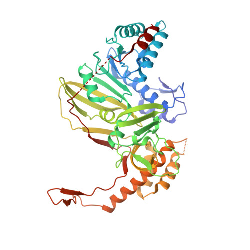
    Positive-sense single-stranded RNA viruses, such as coronaviruses, flaviviruses and alphaviruses, carry out transcription and replication inside virus-induced membranous organelles within host cells 1-7 . The remodelling of the host-cell membranes for the formation of these organelles is coupled to the membrane association of viral replication complexes and to RNA synthesis. These viral niches allow for the concentration of metabolites and proteins for the synthesis of viral RNA, and prevent the detection of this RNA by the cellular innate immune system 8 . Here we present the cryo-electron microscopy structure of non-structural protein 1 (nsP1) of the alphavirus chikungunya virus, which is responsible for RNA capping and membrane binding of the viral replication machinery. The structure shows the enzyme in its active form, assembled in a monotopic membrane-associated dodecameric ring. The structure reveals the structural basis of the coupling between membrane binding, oligomerization and allosteric activation of the capping enzyme. The stoichiometry-with 12 active sites in a single complex-redefines viral replication complexes as RNA synthesis reactors. The ring shape of the complex implies it has a role in controlling access to the viral organelle and ensuring the exit of properly capped viral RNA. Our results provide high-resolution information about the membrane association of the replication machinery of positive-sense single-stranded RNA viruses, and open up avenues for the further characterization of viral replication on cell membranes and the generation of antiviral agents.


  • Organizational Affiliation

    Aix-Marseille Université, CNRS, AFMB UMR 7257, Marseille, France.


Macromolecules
Find similar proteins by:  (by identity cutoff)  |  3D Structure
Entity ID: 1
MoleculeChains Sequence LengthOrganismDetailsImage
Polyprotein P1234472Chikungunya virus strain S27-African prototypeMutation(s): 0 
EC: 2.1.1 (PDB Primary Data), 2.7.7 (PDB Primary Data), 3.1.3.33 (PDB Primary Data), 3.4.22 (PDB Primary Data), 3.6.1.15 (PDB Primary Data), 3.6.4.13 (PDB Primary Data), 3.1.3.84 (PDB Primary Data), 2.7.7.19 (PDB Primary Data), 2.7.7.48 (PDB Primary Data)
UniProt
Find proteins for Q8JUX6 (Chikungunya virus (strain S27-African prototype))
Explore Q8JUX6 
Go to UniProtKB:  Q8JUX6
Entity Groups  
Sequence Clusters30% Identity50% Identity70% Identity90% Identity95% Identity100% Identity
UniProt GroupQ8JUX6
Sequence Annotations
Expand
  • Reference Sequence
Small Molecules
Ligands 1 Unique
IDChains Name / Formula / InChI Key2D Diagram3D Interactions
ZN
Query on ZN

Download Ideal Coordinates CCD File 
M [auth A]
N [auth C]
O [auth E]
P [auth G]
Q [auth I]
M [auth A],
N [auth C],
O [auth E],
P [auth G],
Q [auth I],
R [auth K],
S [auth M],
T [auth O],
U [auth Q],
V [auth S],
W [auth V],
X
ZINC ION
Zn
PTFCDOFLOPIGGS-UHFFFAOYSA-N
Experimental Data & Validation

Experimental Data

  • Method: ELECTRON MICROSCOPY
  • Resolution: 2.60 Å
  • Aggregation State: PARTICLE 
  • Reconstruction Method: SINGLE PARTICLE 
EM Software:
TaskSoftware PackageVersion
MODEL REFINEMENTPHENIX1.17.1
RECONSTRUCTIONRELION3.0.8

Structure Validation

View Full Validation Report


Entry History & Funding Information

Deposition Data


Funding OrganizationLocationGrant Number
ATIP-AvenirFrance--

Revision History  (Full details and data files)

  • Version 1.0: 2020-10-14
    Type: Initial release
  • Version 1.1: 2020-10-21
    Changes: Database references
  • Version 1.2: 2020-12-30
    Changes: Database references
  • Version 1.3: 2021-02-10
    Changes: Database references