6YUZ

Homodimeric structure of the rBAT complex


Experimental Data Snapshot

  • Method: ELECTRON MICROSCOPY
  • Resolution: 2.80 Å
  • Aggregation State: PARTICLE 
  • Reconstruction Method: SINGLE PARTICLE 

wwPDB Validation   3D Report Full Report


This is version 1.0 of the entry. See complete history


Literature

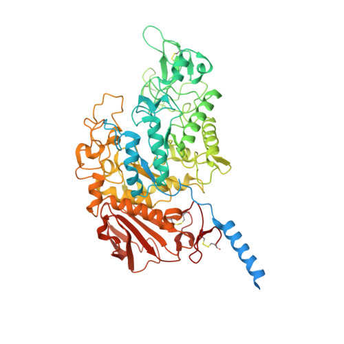
Structural basis for amino acid exchange by a human heteromeric amino acid transporter.

Wu, D.Grund, T.N.Welsch, S.Mills, D.J.Michel, M.Safarian, S.Michel, H.

(2020) Proc Natl Acad Sci U S A 117: 21281-21287

  • DOI: https://doi.org/10.1073/pnas.2008111117
  • Primary Citation of Related Structures:  
    6YUP, 6YUZ, 6YV1

  • PubMed Abstract: 

    Heteromeric amino acid transporters (HATs) comprise a group of membrane proteins that belong to the solute carrier (SLC) superfamily. They are formed by two different protein components: a light chain subunit from an SLC7 family member and a heavy chain subunit from the SLC3 family. The light chain constitutes the transport subunit whereas the heavy chain mediates trafficking to the plasma membrane and maturation of the functional complex. Mutation, malfunction, and dysregulation of HATs are associated with a wide range of pathologies or represent the direct cause of inherited and acquired disorders. Here we report the cryogenic electron microscopy structure of the neutral and basic amino acid transport complex (b [0,+] AT1-rBAT) which reveals a heterotetrameric protein assembly composed of two heavy and light chain subunits, respectively. The previously uncharacterized interaction between two HAT units is mediated via dimerization of the heavy chain subunits and does not include participation of the light chain subunits. The b (0,+) AT1 transporter adopts a LeuT fold and is captured in an inward-facing conformation. We identify an amino-acid-binding pocket that is formed by transmembrane helices 1, 6, and 10 and conserved among SLC7 transporters.


  • Organizational Affiliation

    Department of Molecular Membrane Biology, Max Planck Institute of Biophysics, D-60438 Frankfurt am Main, Germany.


Macromolecules
Find similar proteins by:  (by identity cutoff)  |  3D Structure
Entity ID: 1
MoleculeChains Sequence LengthOrganismDetailsImage
Neutral and basic amino acid transport protein rBATA,
B [auth C]
685Homo sapiensMutation(s): 0 
Gene Names: SLC3A1RBAT
Membrane Entity: Yes 
UniProt & NIH Common Fund Data Resources
Find proteins for Q07837 (Homo sapiens)
Explore Q07837 
Go to UniProtKB:  Q07837
PHAROS:  Q07837
GTEx:  ENSG00000138079 
Entity Groups  
Sequence Clusters30% Identity50% Identity70% Identity90% Identity95% Identity100% Identity
UniProt GroupQ07837
Sequence Annotations
Expand
  • Reference Sequence
Small Molecules
Ligands 2 Unique
IDChains Name / Formula / InChI Key2D Diagram3D Interactions
NAG
Query on NAG

Download Ideal Coordinates CCD File 
C [auth A]
D [auth A]
E [auth A]
F [auth A]
H [auth C]
C [auth A],
D [auth A],
E [auth A],
F [auth A],
H [auth C],
I [auth C],
J [auth C],
K [auth C]
2-acetamido-2-deoxy-beta-D-glucopyranose
C8 H15 N O6
OVRNDRQMDRJTHS-FMDGEEDCSA-N
CA
Query on CA

Download Ideal Coordinates CCD File 
G [auth A],
L [auth C]
CALCIUM ION
Ca
BHPQYMZQTOCNFJ-UHFFFAOYSA-N
Experimental Data & Validation

Experimental Data

  • Method: ELECTRON MICROSCOPY
  • Resolution: 2.80 Å
  • Aggregation State: PARTICLE 
  • Reconstruction Method: SINGLE PARTICLE 
EM Software:
TaskSoftware PackageVersion
MODEL REFINEMENTPHENIXdev-3689
RECONSTRUCTIONRELION3.1

Structure Validation

View Full Validation Report


Entry History & Funding Information

Deposition Data


Funding OrganizationLocationGrant Number
German Federal Ministry for Education and ResearchGermany--
Max Planck SocietyGermany--

Revision History  (Full details and data files)

  • Version 1.0: 2021-01-20
    Type: Initial release