The structural and functional characterization of Malus domestica double bond reductase MdDBR provides insights towards the identification of its substrates.
Caliandro, R., Polsinelli, I., Demitri, N., Musiani, F., Martens, S., Benini, S.(2021) Int J Biol Macromol 171: 89-99
- PubMed: 33412202
- DOI: https://doi.org/10.1016/j.ijbiomac.2020.12.190
- Primary Citation of Related Structures:
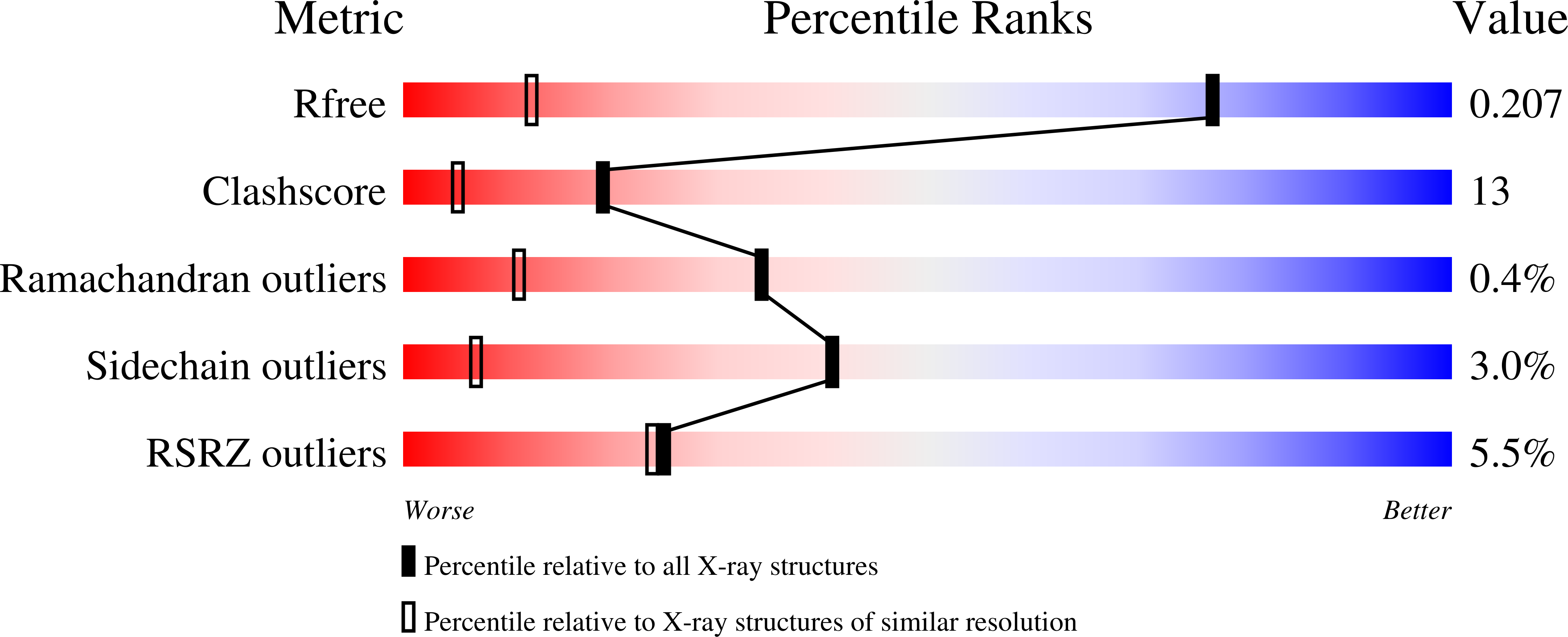
6YSB, 6YTZ, 6YUX - PubMed Abstract:
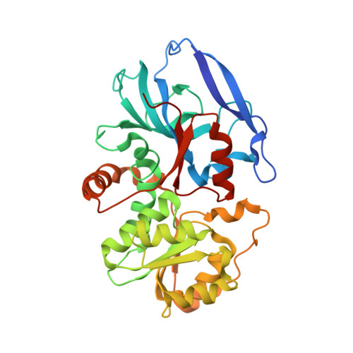
In this study we describe the crystal structures of the apoform, the binary and the ternary complexes of a double bond reductase from Malus domestica L. (MdDBR) and explore a range of potential substrates. The overall fold of MdDBR is similar to that of the medium chain reductase/dehydrogenase/zinc-dependent alcohol dehydrogenase-like family. Structural comparison of MdDBR with Arabidopsis thaliana DBR (AtDBR), Nicotiana tabacum DBR (NtDBR) and Rubus idaeus DBR (RiDBR) allowed the identification of key amino acids involved in cofactor and ligands binding and shed light on how these residues may guide the orientation of the substrates. The enzyme kinetic for the substrate trans-4-phenylbuten-2-one has been analyzed, and MdDBR activity towards a variety of substrates was tested. This enzyme has been reported to be involved in the phenylpropanoid pathway where it would catalyze the NADPH-dependent reduction of the α, β-unsaturated double bond of carbonyl metabolites. Our study provides new data towards the identification of MdDBR natural substrate and the biosynthetic pathway where it belongs. Furthermore, the originally proposed involvement in dihydrochalcone biosynthesis in apple must be questioned.
Organizational Affiliation:
Bioorganic Chemistry and Bio-Crystallography laboratory (B(2)Cl), Faculty of Science and Technology, Free University of Bolzano, Piazza Università 5, 39100 Bolzano, Italy.